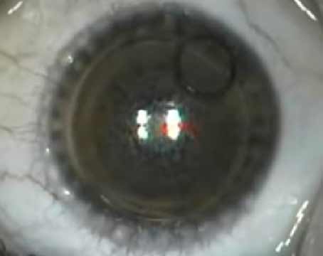
When femtosecond lasers for cataract surgery were first introduced in the United States, their ability to make accurate astigmatic keratotomy or limbal relaxing incisions was front and center, mainly because that was the only way surgeons could charge for the use of the devices. Though surgeons can now charge for more than just the AK, the lasers’ ability to make good, reproducible incisions is no less important, and some surgeons say these incisions may have the most direct impact on postop visual acuity of any aspect of the lasers. In this article, several experienced cataract surgeons share their tips for getting the best results with femtosecond astigmatic incisions.
The Transition from Blades
Surgeons venturing into the world of femtosecond astigmatic keratotomy with their new cataract femtosecond lasers have had to write the rules as they went along, using previous nomograms for manual AK as starting points.
Surgeons say their experience in making the switch from manual to femtosecond arcuate incisions shows how important it is for each surgeon to develop his or her own nomogram based on a series of cases, since some surgeons have experienced very different results in comparison to their blade surgeries. “Limbal relaxing incisions and AK incisions with lasers are definitely a work in progress,” says Draper, Utah, surgeon Robert Rivera, who uses both the Optimedica Catalys and the Alcon LenSx lasers. “Now that we have the laser’s consistency and precision from case to case, we can start to assess other factors in the corneal incisions to a degree we couldn’t before, such as the exact length of the incision arc, its depth and even the optical zone. We can start to look at these in a more scientific way because we’ve taken that ability out of the hand and placed it on the computer’s motherboard, if you will. You have to be careful and not make the assumption that the manual nomogram will give you the same result with the femtosecond laser as it would with a diamond blade; the tendency, in my experience, has been toward a little overcorrection.”
Nashville surgeon Ming Wang, though, has had the opposite experience. “I found that, once I had the WaveTec Optiwave Refractive Analysis intraoperative wavefront system to intraoperatively check the total refractive astigmatism, almost universally the laser AK/LRIs are undercorrecting,” he says. “This is a systematic error—not a random error—and was previously unknown prior to the availability of the ORA. Furthermore, given the fact that the astigmatic corrective effect of the laser AK/LRI incisional keratotomy will be reduced postoperatively due to the filling in of the incisions, there will be even more of an undercorrection.”
With these tendencies toward over- or undercorrection, surgeons say their approaches to astigmatic correction can be used simply as starting points for others to hone their nomograms. It’s important to note that, in their approaches, several surgeons refer to the Donnenfeld and the Nichamin LRI nomograms, which are part of a manual, blade LRI planning program provided by Abbott Medical Optics at lricalculator.com. Many surgeons use these blade nomograms as jumping off points for their laser nomograms. Here are the steps some of them take.
“What we’ve been using for femtosecond incisions is the Donnenfeld nomogram for manual LRIs that’s based on an 11-mm optical zone,” explains Long Island surgeon Eric Donnenfeld. “But we decrease the nomogram by 33 percent and reduce the optical zone to 9 mm. So, if the nomogram says to treat with a 60-degree arc, I use a 40-degree arc. With this, we get about a 70 percent reduction in the cylinder.”
“I basically use the Donnenfeld nomogram, but the difference between the manual and laser is that, for the laser, I program it for 80 percent of the stromal thickness, and I perform it at a 9-mm optical zone,” explains Dr. Wang. “I also vary the number of incisions between a single incision and paired incisions depending on the location of the primary and secondary wounds for the cataract surgery itself.”
In developing an approach that worked for the IntraLase laser, Tampa, Fla., surgeon T. Hunter Newsom says you can find success altering both the blade nomogram and the laser’s power output. He uses the anterior side-cut function of the Intralase-enabled keratoplasty software. “We use Dr. Nichamin’s NAPA nomogram from the AMO LRI calculator website,” he says. “We approached it like a blade surgery’s nomogram and then modified it, moving the optical zone in a little bit. Rather than trying to reduce our nomogram, we wanted to leave it as it was and adjust the energy of the laser.
“When you use a femtosecond laser to make an incision, it makes a series of spherical spots next to each other, similar to the edge of a postage stamp or the holes in the pages of a spiral notebook,” he explains. “You can connect these holes tightly enough so it produces an effect that’s the same as having a bladed incision that’s wide open. You can adjust this process, however, by altering the spot size and the energy. So, say we have a 3-µm spot separated from the next spot by 3 µm at 10 mJ of energy. The resulting air bubble won’t be 3 µm in size, but will instead be 5 µm because the explosion is so large the air bubble will separate more tissue. This will result in incisions that flop open on their own, just as if you’d used a diamond blade. We didn’t want that. So, we were able to turn the laser energy down to 1 mJ but keep a 3 µm spot size, creating a weaker shot. This creates a spot of about 2.5 µm, so as you move over by 3 µm, the spots never really connect. This reduces the effectiveness of the incision you’re making versus a blade but it makes the incision tighter, so it won’t open immediately. With this method, we can just program the treatment using the NAPA nomogram and instead of reducing it by 30 percent, we just reduce the laser energy to reach a point where the procedure gives us the equivalent of a 10-mm blade incision at full thickness.”
To see how his approach compared to blade incisions, Dr. Newsom performed a six-month chart review of 62 eyes of 45 patients who received premium IOLs and who required astigmatic corrections ranging from 0.75 to 3.1 D. The mean attempted cylinder correction was 1.32 D (r: 0.75 to 3.1) for the femtosecond incision group and 1.13 D (r: 0.75 to 1.75) for the blade LRI group. Postop, residual manifest cylinder was 0.29 D (r: 0.0 to 0.75, ±0.29) in the femtosecond group and 0.42 D (r: 0.0 to 1.25, ±0.38) in the blade group. Dr. Newsom says this is a statistically significant difference in achieved correction (p=0.047), with 78 percent for the femtosecond and 63 percent for the blade. The mean visual acuity postop was 20/27 for FAI and 20/28 for LRI. Dr. Newsom points out that, though the femtosecond procedure decreased astigmatism by a greater amount, the blade incision was excellent, as well, doing a good job at getting patients below 0.5 D of astigmatism.
As surgeons learn more about the effects of femtosecond incisions, they say different wound architectures may emerge that yield even better results. “In the past, we assumed an incision perpendicular to the cornea was best,” says Dr. Rivera. “But, at some point, we may look into new wound architectures to see if we can achieve better or tighter astigmatic corrections. Several years ago, Richard Mackool discussed his idea for a penetrating LRI technique in which he’d take his cataract surgery blade and use it to create a full-thickness penetrating incision contralaterally to the steep axis of cylinder. In that case, the incision was basically as close to a mirror image of the primary cataract wound as possible, with full-thickness penetration. So that would be another way to correct astigmatism, and I wouldn’t be surprised if, at some point down the road, we start modifying the wound architecture to see if other types of incisions—be they tunnels, corneal pockets or things of that sort—may not give us more stability or accuracy overall.”
Titrating the Incisions
One aspect of laser AK that surgeons can use to their advantage in the search for better outcomes is the fact that these incisions stay closed until the surgeon opens them, minimizing their initial effect. Physicians say this titratability helps increase the margin for error as they hone their femtosecond AK nomograms.
“The way the femtosecond laser creates the incisions allows you to treat astigmatism with the incision later,” says Dr. Rivera. “Many of us leave the incision or incisions unopened at the time of surgery, and then, depending on what the patient’s postop refraction is, we go in and titrate the correction by opening the incisions at the slit lamp. Most of the time, I do paired astigmatic incisions, though in some cases I will place my primary cataract wound at the patient’s steep axis. If someone has 1.5 D of cylinder, for example, I’ll operate on the steep axis with a 2.9-mm entry wound—my cataract wound typically corrects
0.5 D of cylinder—and I’ll then place a 30 to 40 degree contralateral incision that I leave untouched at the time of surgery. Two weeks postoperatively, I’ll see what the patient’s refraction is and consider opening the contralateral incision to achieve more cylinder control. So, by pairing my entry incision with an LRI, I can typically take care of 1.5 to 2 D. Beyond that level of astigmatism we begin looking at toric intraocular lenses, surface refractive surgery or LASIK.
“The opening procedure is performed right at the slit lamp,” Dr. Rivera continues. “I’ve actually been surprised in a number of cases in which the patient didn’t require the incision to be opened at all. Having the luxury of leaving the incisions untouched depending on the level of the postoperative cylinder is a nice benefit of femtosecond astigmatic incisions.” Dr. Rivera currently makes his incisions at the 9 mm optical zone.
Dr. Newsom also will open his wounds postop to titrate the effect. “Postop, the wounds can still be opened without difficulty,” he says. “If we didn’t get all the astigmatism we wanted by making the incisions, we can go back and open them up a little at a week, two weeks or a month postop. In terms of the range of correction, we can get 2 D and below.”
Dr. Wang works ORA technology into the mix to titrate the astigmatic incisions intraoperatively. “First, I make the laser AK incision, but I don’t open it,” he says. “For the incision, I use the Donnenfeld nomogram with 80 percent corneal depth at a 9 mm optical zone. Once the IOL is implanted, I perform ORA intraoperative wavefront aberrometry, then titrate the amount of astigmatism treatment by opening the AK incision based on ORA readings. When the patient leaves the operating room, the astigmatism is treated.
“We’ve conducted a study of 96 consecutive eyes comparing intraoperative cylinder measured by ORA with the postoperative refractive cylinder at one month and three months,” Dr. Wang continues. “We found that the mean difference between the magnitudes of cylinders measured intraoperatively by ORA and those measured postoperatively were 0.64 ±0.44 D at one month and 0.66 ±0.44 D at three months, with vector centroid and deviation of 0.24 ±0.52 D at 138 degrees at one month and 0.029 ±0.54 D at 140 degrees at three months. We’ve thus shown that ORA provides a good predictability in magnitude and vector of cylinder, and we will be presenting this study at the upcoming ASCRS meeting in San Francisco.”
The Intrastromal Route
If femtosecond astigmatic incisions are cutting edge, then femtosecond intrastromal incisions—which don’t break the epithelium—are on the bleeding edge of technology. Exact nomograms for these incisions are still being ironed out, though surgeons have initial results they can share.
“A full-penetrating femtosecond astigmatic incision is conceptually similar to a manual LRI, depending on the energy setting of the laser,” says San Diego surgeon Steven Schallhorn, MD, who is developing an intrastromal nomogram. “But an intrastromal incision is different because you don’t break Bowman’s layer. Ultimately, the arc angle is a little longer or the optical zone is a little smaller; we use a 7-mm optical zone. You get more effect with a smaller optical zone, and, of course, more effect with a larger cut angle. Those are the two adjustments we’ve made.” Dr. Schallhorn says the IntraLase is programmed so that the anterior depth keeps the incision below Bowman’s layer.
To give a taste of where the nomogram stands, Dr. Schallhorn recounts a recent patient who was treated with intrastromal AK. “This patient was an accountant who had undergone a refractive lens exchange and had 1 D of residual astigmatism that was causing her visual symptoms,” he explains. “Since the spherical equivalent in that eye was plano, this was a perfect opportunity to perform a femtosecond AK. So, three months after the RLE she underwent intrastromal AK, receiving two symmetrical 45-degree arcs at the 7-mm zone and 80 percent depth. Postop, her uncorrected vision was 20/16 and she went on to have an RLE procedure in the other eye.” Dr. Schallhorn says the intrastromal upper limit is 2 D, which would require two 75-degree arcs.
“We’re in the process of refining our nomogram after having performed approximately 150 cases,” Dr. Schallhorn says. “Once it’s refined, we will undertake a clinical trial to validate the nomogram. This will all take place within the next several months.”
Dr. Newsom says he has tried intrastromal incisions but warns that they can pose challenges. “We found that when we used the standard femtosecond energy settings for an intrastromal incision, we started to get some microperforations, where the incision would go through Descemet’s and the endothelium,” Dr. Newsom says. “Also, since the incision is going only through the stroma and not Bowman’s, you have to make a much longer incision to achieve the same effect as a full-penetrating incision. So, instead of making a two clock hour incision to reduce a diopter of astigmatism, you may have to make a four clock hour incision. You really have to look at these intrastromal incisions one hour after surgery, such as with an anterior chamber OCT, otherwise you won’t notice these microperforations. We noticed that we were getting more of these microperforations than we thought, and that’s when we started decreasing the energy until we didn’t get them. But then we said, we’re so worried about going into the anterior chamber, why don’t we just break the surface with the incision and stick with the full-penetrating incisions?”
In terms of his intrastromal experience, Dr. Schallhorn says he hasn’t had a problem with microperforations. “We haven’t observed any anterior chamber bubbles or problems with Descemet’s,” he avers. “We use the Pentacam to diligently analyze the corneal thickness, and set the posterior depth to 80 percent of the thinnest cornea over the planned incision sites.”
Even though intrastromal nomograms are still being refined, Dr. Schallhorn has analyzed the procedure’s accuracy so far. “We’re seeing about a 50-percent reduction in the absolute value of the astigmatism,” he says. “When you look at the correction ratio, which takes into account the meridian of the astigmatism in addition to the value, we’re getting about 75 percent of the effect we want, meaning undercorrection. Even though we can still improve on this, you have to remember that this is right out of the box with the laser. We’re currently analyzing our data to refine the nomogram and improve the results.”
Dr. Rivera says he thinks the precision of the femtosecond laser holds great promise for the future of astigmatic incisions. “When we target plano in an astigmatic patient, we’re typically getting that about 90 to 95 percent of the time with little residual astigmatism,” he says. “I’ve had to bring a couple of these patients back in order to perform an enhancement for astigmatism, but the procedure is getting better all the time.” REVIEW


