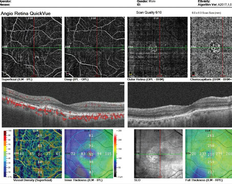
For patients with macula-off primary rhegmatogenous retinal detachments, scleral buckling may be the wiser repair approach, according to a recent study published in BMC Ophthalmology. At present, primary RRDs are typically addressed using scleral buckling, pars plana vitrectomy, combined scleral buckling and pars plana vitrectomy, and pneumatic retinopexy. However, despite successful repair, it’s not uncommon for some eyes to go on to develop complications such as cystoid macular edema or epiretinal membrane. The study authors investigated and compared incidence rates and risk factors associated with CME and ERM after primary RRD repair with scleral buckling and pars plana vitrectomy. They reported that macular status and repair approach play a role in risk.
The retrospective observational cohort study included 62 consecutive patients with primary RRD who were treated with either scleral buckling or pars plana vitrectomy. Those who underwent scleral buckling were young, phakic patients without posterior vitreous detachment, high myopic patients and those whose RRD was associated with anterior or interior retinal tears.
Patients who underwent pars plana vitrectomy were pseudophakic or had media opacity and posterior breaks precluding scleral buckling use. Macular changes were evaluated at the three- and six-month postop visits. For phakic patients whose media opacity or lens bulging hindered surgical maneuvers, phacoemulsification and IOL implantation was also performed.
Inner limiting membrane peeling, a non-standard surgical procedure for RRD repair but one that’s been reported to confer greater macular elasticity during reattachment, was performed randomly in the macula-off (15/30 patients) and the macula-on RRD “pending foveal detachment” (2/4 patients) subgroup.
Study co-author Matteo Ripa, MD, of the Department of Ophthalmology, William Harvey Hospital, East Kent Hospitals University NHS Foundation Trust in the United Kingdom, explains that surgeons should consider macular status to be a critical factor requiring constant evaluation in “primary retinal detachment repair management due to its role in determining final visual and functional outcomes.” He says, “Assessing the risk factors and incidence of ERM and CME formation after scleral buckling and pars plana vitrectomy in patients who developed primary RRD, we found that macula-off status significantly increased the risk of CME by odds-ratio (OR)=4.3 times compared with macula-on, regardless of the procedure (p=0.04), whereas neither the macula-off status in patients who underwent pars plana vitrectomy nor the ILM peeling significantly increased the risk of postoperative CME (OR=1.73, p=0.4 and OR=1.8, p=0.37, respectively).
“Furthermore, our results clearly show significant differences in CME incidence when comparing patients who underwent pars plana vitrectomy and scleral buckling (i.e., 33 percent of patients (14/42) who underwent pars plana vitrectomy developed a postoperative CME, and no CME cases were found in the scleral buckling group, p=0.001),” he continues. “At the end of the follow-up, resolution of CME was observed in 13 out of 14 patients (92.86 percent). Despite the treatment (indomethacin three times daily up to resolution), CME didn’t resolve in only one patient.
“Regarding the OCT CME morphology, we mostly found central cystoid spaces within the inner or outer retina ± subretinal fluid with no diffuse macular distribution,” he says. “Furthermore, according to the CME morphology, only six cases of CME were associated with ERM. Several factors could have been implied in the CME genesis, such as inflammation, tractions and macular status. Nonetheless, inflammation played a crucial role, as eight out of 14 (57.14 percent) cases of CME weren’t associated with ERM (a possible additional tractional mechanism).”
When asked about phacovitrectomy for RRD repair, Dr. Ripa noted that this combined procedure has several advantages but may be unsuitable for certain patients. He explains, “First, in patients with significant cataracts, a combined procedure in which the surgeon addresses the cataract first optimizes the view and surgical access to the retina, thus improving the visualization for more detailed retinal work. Second, it leads to an overall faster recovery time as pars plana vitrectomy can induce lens opacification that’s most likely to occur in a reasonably short time, thus affecting postoperative visual recovery. Third, it eases surgical maneuvers reducing the so-called ‘lens touch’ that may lead to increased complication rates in subsequent cataract surgery. Moreover, despite a high risk of ‘refractive surprise,’ many surgeons remove the natural lens in combination with the pars plana vitrectomy, regardless of the cataract.
“Despite these several advantages, the higher risk of CME after combined surgeries cannot be underrated, as the postoperative inflammation can compromise functional recovery,” he says. “Therefore, according to the study results, every surgeon should balance the benefits and risks to properly manage primary retinal detachment repair using a more personalized therapeutic approach.”
Dr. Ripa points out that study limitations included its retrospective, non-randomized nature and small sample size (62 patients, 20 in scleral buckling, and 42 in pars plana vitrectomy subgroups); that it didn’t “consider a multivariate analysis of several risk factors for CME and the ERM development after primary RRD repairs, such as age, extensive vs. not extensive use of endolaser retinopexy or cryotherapy, type of tamponade used, possible additional surgical maneuvers, number of previous surgeries and RRD surgery duration. In addition, the retinal tears numbers and their location weren’t considered as a deciding factor for the surgical technique adopted”; that “ILM peeling was performed on macula-off and macula-on ‘pending foveal detachment’ but not in macula-on ‘properly so-called’ ”; and that “six-month outcomes may not necessarily indicate long-term outcomes, as ERM and CME may arise long after a successful primary RRD repair.”
Overall, he advises surgeons to consider macular status when approaching primary retinal detachment repair since this factor had such a significant effect on postoperative complications, independent of surgical technique. He says, “Scleral buckling may be less likely to be related to postoperative surgical complications than pars plana vitrectomy in achieving surgical primary RRD repair, according to other research that reported a higher risk of CME associated with any ab-interno macular surgery.”
Physicians Deal with Corneal Infections from Artificial Tears
Amid a nationwide recall of Global Pharma Health Care’s artificial tears (sold under the names EzriCare and Delsam Pharma) due to the products’ possible contamination, ophthalmologists at Bascom Palmer published a report on a patient whose infection may be linked to the agent1. As of mid-March, according to the Centers for Disease Control and Prevention, 68 patients in 16 states have been infected with a rare strain of extensively drug-resistant P. aeruginosa. Three patients have died and there have been eight reports of vision loss and four enucleations due to the infections. The CDC adds that isolates were identified from cultures of sputum or bronchial wash (15), cornea (17), urine (10), other nonsterile sources (4), blood (2), and from rectal swabs (26). Some patients had specimens collected from more than one site.
In the study, the researchers recount how an older man presented with complaints of right eye pain and decreased vision that had lasted for the past day. His medical history included coronary artery disease, diabetes and chronic obstructive pulmonary disease. He wore contact lenses but denied sleeping in them or overuse. He also reported the use of EzriCare artificial tears. His best-corrected visual acuity was hand motion in the right eye and 20/20 in the left. Intraocular pressures were 29 mmHg in the right eye and 14 mm Hg in the left. In the patient’s right eye, the physician noted conjunctival hyperemia, a 6 × 5-mm corneal infiltrate with overlying epithelial defect, and 2-mm hypopyon. Ultrasound was normal without membranes or vitritis.
The authors say that, since there’s currently a rash of multi-drug resistant infections due to the use of EzriCare drops and a recent CDC warning about the situation, they treated the eye with with topical fortified vancomycin, fortified tobramycin and trimethoprim-polymyxin drops every hour while awake. They cultured both the infiltrate and the EzriCare artificial tears. The corneal culture was positive for P. aeruginosa with high resistance to fluoroquinolones; aminoglycosides, including amikacin and tobramycin; and cephalosporins, with moderate carbapenem resistance (minimum inhibitory concentration = 4). The EzriCare culture was also positive for P. aeruginosa resistant to fluoroquinolones, aminoglycosides, and cephalosporins, with higher carbapenem resistance (minimum inhibitory concentration = 8). Based on the bacterial sensitivities, they say that the patient was continued on trimethoprim-polymyxin every hour and switched to imipenem-cilastatin every two hours, “as this antibiotic class had the lowest resistance of those tested.” The patient is currently undergoing treatment with close monitoring, as he had persistent infection and vision loss at his last follow-up, the researchers say.
In a commentary on the outbreak in JAMA Ophthalmology, Kathryn Kolby, MD, PhD, chair of the department of Ophthalmology at the NYU Grossman School of Medicine, writes, “… the current outbreak of Verona Integron-mediated Metallo-ß-lactamase (VIM) and Guiana-Extended Spectrum-ß-Lactamase (GES)-producing carbapenem-resistant (VIM-GES-CRPA), a rare strain of extensively drug-resistant Pseudomonas aeruginosa, associated with the use of carboxymethylcellulose sodium (EzriCare) multidose preservative-free artificial tears may be a wake-up call for the field. … This outbreak is a harsh reminder that all eye drops, including artificial tears, are medications with potential adverse effects, most commonly ocular but potentially systemic.”
1. Shoji MK, Gutkind NE, Meyer BI, et al. Multidrug-Resistant Pseudomonas aeruginosa Keratitis Associated With Artificial Tear Use. JAMA Ophthalmol. March 22, 2023 (online article).
Industry News Aurion Receives Approval In Japan Aurion Biotech received regulatory approval from Japan’s Pharmaceuticals and Medical Devices Agency for its novel cell therapy, Vyznova, for the treatment of bullous keratopathy of the cornea. The company believes this is the first-ever regulatory approval in the world for an allogeneic cell therapy to treat corneal endothelial disease. The breakthrough innovation of this cell therapy is to enable fully differentiated corneal endothelial cells to regenerate outside the body, the company says. Healthy cells from a donor cornea are cultured in a novel, multi-step, proprietary and patented process that produces off-the-shelf, allogeneic, fully differentiated CECs. The endothelial cells are then injected intracamerally where they re-populate into a healthy mono-layer and start removing fluid from the cornea, thereby decreasing corneal edema, the company says.
Aviceda Therapeutics submitted to the Food and Drug Administration an Investigational New Drug application for its lead intravitreal ocular asset, AVD-104 (a novel glycan-coated nanoparticle) to treat geographic atrophy secondary to AMD.
Iveric Bio Gets Priority Review Iveric Bio announced the Food and Drug Administration completed its filing review and accepted the company’s New Drug Application for avacincaptad pegol, a novel investigational complement C5 inhibitor for the treatment of geographic atrophy secondary to age-related macular degeneration. The New Drug Application, based on efficacy and safety results from the GATHER1 and GATHER2 clinical trials, was granted Priority Review with a Prescription Drug User Fee Act goal date of August 19. |


