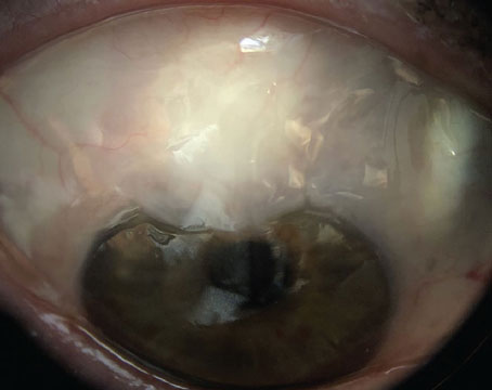
Investigators assessed the recurrence rate of active macular neovascularization in patients with neovascular age-related macular degeneration previously followed in a treat-and-extend regimen in which treatment had been stopped due to disease stability, as part of a prospective cohort study.
A total of 105 nAMD patients previously followed in a treat-and-extend regimen with aflibercept injections were included. All patients with a dry macula on three consecutive visits 12 weeks apart were eligible to participate. Patients were examined at baseline, and then monitored for disease recurrence four, six, eight, 10 and 12 months after the last injection.
Main outcome measures included the proportion of patients with recurrent disease within 12 months after the last injection, and BCVA change at the time of recurrence and after resumed therapy.
Here are some of the findings:
- Evidence of recurrent nAMD was seen in 54/102 patients (52.9 percent) after 12 months of follow-up.
- The mean time to recurrence after the last injection was 6.7 ±2.2 months. The best-corrected visual acuity decreased from 71.7 ±10
ETDRS letters at baseline to 68.1 ±11.1 letters at the recurrence (p=0.12). - After treatment was resumed, BCVA increased to 71.4 ±10 letters (p=NS compared to baseline).
- Patients with a pigment epithelial detachment at baseline had a 74 percent (14/19) recurrence rate compared to 48 percent (40/83) in subjects without a PED (p<0.05).
- Only 22/54 (40.7 percent) of the patients with recurrent disease had symptoms of visual loss or metamorphopsia.
Investigators reported that recurrent nAMD was common in previously stable patients when anti-VEGF injections were suspended. They wrote that it was difficult to predict which patients would have a recurrence, as most didn’t have symptoms in the early stages of reactivation. Investigators stressed the importance of long-term follow-up and noted that early detection of recurrent disease can improve the chances of maintained visual function.
Ophthalmol Retina 2021; Mar 25 [Epub ahead of print].
Aslanis S, Amrén U, Lindberg C, et al.
OCTA Artifacts in Glaucoma
Researchers determined the prevalence of different artifacts on optical coherence tomography angiography images of healthy and glaucomatous eyes, and evaluated the characteristics associated with the increased likelihood of obtaining poor quality images.
A total of 649 eyes of 368 healthy individuals, glaucoma suspects and glaucoma patients were included.
Angiovue high-density and non-HD optic nerve head and macula OCTA images of participants were evaluated by four expert reviewers for the presence of different artifacts, including: eye movement; defocus; shadow; decentration; segmentation error; blink; and Z offset in the superficial vascular layer. Each OCTA scan was designated as good or poor quality based on the presence of artifacts. Researchers evaluated the association of demographic and ocular characteristics with the likelihood of obtaining poor-quality OCTA images, using a generalized linear mixed model.
Main outcome measures included the prevalence of OCTA artifacts and factors associated with increased likelihood of capturing poor-quality OCTA images.
A total of 5,263 OCTA images were evaluated. Here are some of the findings:
- Overall, 33.9 percent of the OCTA images had poor quality.
- The majority of images with acceptable quality scores (QS ≥4) had no artifacts (76.6 percent).
- Other images had one (13.6 percent), or two or more artifacts (9.8 percent).
- Older age (p<0.001), male gender (p=0.045), worse visual field mean deviation (p<0.001), absence of eye tracking (p<0.001) and macular scan area (p<0.001) were associated with a higher likelihood of obtaining poor-quality images.
- In images with acceptable quality scores, commercially available quality measures, including quality scores and signal strength index, had area under the ROC curves of 0.65 and 0.70, respectively, detecting good-quality images.
Researchers advise that clinicians should conduct a systematic scan review to ensure appropriate interpretation of OCTA images. The investigators add that OCTA images should be reacquired whenever an apparent and correctable artifact is present on the captured image.
Ophthalmol 2021; April 2 [Epub ahead of print].
Kamalipour A, Moghimi S, Hou H, et al.