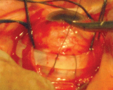
The vitreous plays an integral role in a variety of retinal diseases.1-3 The pathogenic mechanisms by which the vitreous affects the retina are not completely understood, but abnormalities in vitreoretinal adhesion may have a direct effect on retinal oxygenation and on growth factor and cytokine production within the eye. Currently, surgical manipulation via pars plana vitrectomy is used to manage this interface. Recent developments now allow for manipulation of the vitreous without surgical intervention.
“Vitreodynamics” is a term used by Michael T. Trese, MD, in the recognition of changes in vitreous oxygenation and growth factor levels that result from pharmacologically induced changes in vitreous molecular structure.4 The vitreous, with its high water content and relatively few molecular and cellular components, is a unique extracellular matrix. It allows for rapid diffusion of injected agents, making it ideal for pharmacologic manipulation. The predominant macromolecular components of the vitreous are hyaluronan and collagens. Hyaluronan, a disaccharide glycosaminoglycan consisting of glucuronic acid and N-acetyl glycosamine, exists as a highly hydrated series of coiled, long molecular chains arranged in a linear helix.2,5
Vitreous collagens are non-branching fibrils 7 to 28 nm in diameter consisting of covalently linked collagen molecules. Analysis of vitreous reveals collagen type II, a hybrid of type V and type XI, and type IX distributed in a ratio of 75:10:15, respectively. Although the majority of collagen is type II, types V/XI and IX facilitate stability of the vitreous gel while type IX collagen promotes cohesion between the fibrils and hyaluronan. The fibrils are structured with type V/XI at the core, surrounded by type II with type IX on the fibril surface. The assembly of these collagens enhances fibril stability and strength. Interactions between the collagen fibrils and hyaluronan create the unique optical and mechanical properties of the vitreous. Large domains of hyaluronan stabilize and disperse the collagen fibrils, minimizing light scattering. The viscoelastic properties of the vitreous result from complex molecular interactions between the collagen fibrils and hyaluronan as well as other glycosoaminoglycans, such as chondroitin 6-sulfate. These glycosaminoglycans aggregate together, via hydrophobic and hydrogen bonding, to create a fragile, three-dimensional, honeycomb-like structure. This fragility may be exploited by pharmacologic agents.2,6
The mechanical relationship between the vitreous and the retina is mediated by the posterior vitreous cortex and the internal limiting membrane, as the posterior vitreous cortex consists of densely packed collagen fibrils arranged tangentially upon the ILM. The adhesion of the posterior vitreous cortex is a dynamic process with topographic and age-related variations. With no apparent gross anatomic correlates to explain the occurrence of vitreoretinal adhesion, it is believed that mediation of this adhesion is by a molecular glue consisting of glycoproteins, such as fibronectin, laminin and chondroitin, as well as integrins and other glycoconjugates. This glue is an important target in pharmacologic vitreolysis.2,5
As with other extracellular matrixes, the vitreous contains matrix metalloproteinases, which are regulated by tissue inhibitors of MMP (TIMP). MMP and TIMP play both a physiologic and pathologic role in the vitreous, as these enzymes contribute to the proteolytic state that clears the vitreous of potential opacification and maintains optical clarity. In disease states, MMP have been implicated in proliferative vitreoretinopathy, retinopathy of prematurity and diabetic retinopathy.7
| Enzymatic vitreous disruption...is a non-surgical method of inducing a PVD and liquefaction of the vitreous, allowing for a cleaner retinal surface. |
The Pharmacologic Vitreolysis for Vitreomacular Traction (Microplasmin for IntraVitreous Injection-Traction Release without Surgical Treatment) trial is the largest interventional clinic program evaluating the vitreoretinal interface in patients with retinal disorders. Six hundred fifty-two patients from 90 centers across the United States and Europe participated in a Phase III program to evaluate the utility of microplasmin. The MIVI-006 trial was a Phase III study to evaluate a single injection of ocriplasmin for treatment of focal VMA in the United States. The MIVI-007 trial was a Phase III study to evaluate a single injection of ocriplasmin for treatment of focal VMA in the United States and Europe. These trials were designed to provide more definitive information as to the value of microplasmin enzyme in vitreomacular traction.
These Phase III, multicenter, randomized, placebo-controlled, double-masked clinical trials were designed to investigate intravitreal microplasmin 125 µg in eyes with symptomatic focal VMA <400 µm. Exclusion criteria included proliferative retinopathy, macular hole diameter >400µm, high myopia, prior retinal detachment or vitrectomy. Subjects were randomized to microplasmin or placebo intravitreal injection (allocation ratio 3:1). The primary endpoint was non-surgical resolution of VMA at day 28, determined by the Central Reading Center OCT evaluation. Secondary endpoint assessments included total PVD at day 28 (determined by ultrasound), avoidance of vitrectomy, proportion of patients with non-surgical MH closure (determined by reading center), and improvement in visual acuity and quality of life as measured by the VFQ-25, National Eye Institute Visual Functioning Questionnaire. Eyes were followed for six months.
The groups were relatively well-matched in age, gender, ethnicity and baseline vision. There was a higher proportion of patients in the microplasmin-treated group with focal VMA >1,500 µm. A statistically significant improvement in the resolution of VMA was seen in the microplasmin-treated group—26.4 percent compared to 10.2 percent who received a placebo injection (p<0.001) achieved resolution of their VMA at 28 days. Patients without epiretinal membranes were more likely to have resolution of VMA—37.4 percent of the microplasmin-treated patients achieved VMA resolution at 28 days compared to 14.3 percent of placebo patients (p<0.001).
Secondary key endpoints were also evaluated. A total PVD was achieved at day 28 in 13.3 percent of microplasmin-treated patients compared to 3.7 percent in the placebo group, which was statistically significant. In patients with FTMH, 40.6 percent of patients saw closure at 28 days with one injection compared to 10.6 percent of the placebo group (p<0.001), with 40.6 percent with closure at the end of the study in the microplasmin-group versus 17 percent in the placebo group. The need for surgical intervention was significantly less in the microplasmin group (17.6 percent vs. 26.7 percent placebo).
Full-thickness MH patients experienced a statistically significant improvement in vision from baseline. At the end of six months, 23.7 percent of the patients treated with microplasmin achieved at least a 10-letter improvement in vision compared to 11.2 percent of patients in the placebo group, and a 15-letter improvement was seen in 9.7 percent of microplasmin patients compared to 3.7 percent in the placebo group. Overall vision improvement was better in the microplasmin group (28 percent) compared to placebo (17.1 percent), with a 15-letter improvement in the microplasmin group at 12.3 percent compared to 6.4 percent in the control. Quality of life, evaluated via the VFQ-25, was improved in the treated group.
Microplasmin was found to be safe and well-tolerated. Serious and severe adverse events were seen equally in both groups. The adverse events seen more often in the microplasmin group were those associated with PVD induction—vitreous floaters and photopsias. There was no increase in risk for retinal tear or retinal detachment in the microplasmin group compared to placebo. There was also no increase in cataract formation in the microplasmin group. Overall, microplasmin was well-tolerated with less risk of retinal tear or retinal detachment or cataract compared to surgery.
This trial demonstrated that microplasmin successfully resolved vitreomacular adhesion, resolved full-thickness holes without surgical intervention, delivered an improvement in the vision of patients without surgical intervention, and was well-tolerated. Both trials met primary endpoints with high statistical significance. Both trials met PVD induction and statistically significant increase in rate of non-surgical macular hole closure, vision improvement, and improvement on the VFQ-25 questionnaire. Microplasmin may become a new tool in the armamentarium of retinal specialists.
Drs. Faia and Williams practice at Associated Retinal Consultants, William Beaumont Hospital, 3535 West 13 Mile Rd. #344, Royal Oak, MI 48073. Contact Dr. Williams:
GWilliams@beaumont.edu. Dr. Faia has no financial interests to disclose. Dr. Williams is a shareholder and consultant for ThromboGenics Ltd. and holds intellectual property on the use of plasmin.
1. Sebag J. Anomalous posterior vitreous detachment: A unifying concept in vitreoretinal disease. Graefe’s Arch Clin Exp Ophthalmol 2004;242:690-698.
2. Williams G. Pharmacologic manipulation of the vitreous during pars plana vitrectomy. In: Alfaro D, Liggett P, eds. Vitreoretinal Surgery of the Injured Eye. Philadelphia: Raven, 1998:141-150.
3. Krebs I, Brannath W, Glittenberg C, et al. Posterior vitreomacular adhesion: A potential risk factor for exudative age-related macular degeneration? Am J Ophthalmol 2007;144:741-746.
4. Quiram P, Leverenz V, Baker R, et al. Microplasmin-induced posterior vitreous detachment affects vitreous oxygen levels. Retina 2007;27:1090-1096.
5. Sebag J. Anatomy and pathology of the vitreoretinal interface. Eye 1992;6:541-552.
6. Sebag J. Molecular biology of pharmacologic vitreolysis. Trans Am Ophthalmol 2005;103:473-494.
7. Sethi C, Bailey T, Luthert P, Chong N. Matrix metalloproteinase biology applied to vitreoretinal disorders. Br J Ophthalmol 2000;84:654-666.
8. Uemura A, Yamamoto S, Sato E, Sugawara T, et al. Vitrectomy alone versus vitrectomy with simultaneous intravitreal injection of triamcinolone for macular edema associated with branch retinal vein occlusion. Ophthalmic Surg Lasers Imaging. 2009 Jan-Feb;40(1):6-12.
9. Gandorfer A. Enzymatic vitreous disruption. Eye 2008; 22:1273-1277.
10. Trese M. Enzymatic-assisted vitrectomy. Eye 2002;16: 365–368.
11. Gandorfer A, Ulbig M, Kampik A. Plasmin-assisted vitrectomy eliminates cortical vitreous remnants. Eye 2002; 16: 95–97.
12. de Smet MD, Valmaggia C, Zarranz-Ventura J, Willekens B. Microplasmin: Ex vivo characterization of its activity in porcine vitreous. IOVS 2009; 50(2):814-819.
13. Jorge R, Oyamaguchi EK, Cardillo JA, Gobbi A, et al. Intravitreal injection of dispase causes retinal hemorrhages in rabbit and human eyes. Curr Eye Res 2003; 26: 107–112.
14. Frenzel EM, Neely KA, Walsh AW, Cameron JD, Gregerson DS. A new model of proliferative vitreoretinopathy. Invest Ophthalmol Vis Sci 1998; 39: 2157–2164.
15. Gandorfer A, Rohleder M, Sethi C, Eckle D, Welge-Lussen U, Kampik A et al. Posterior vitreous detachment induced by microplasmin. Invest Ophthalmol Vis Sci 2004; 45: 641–647.
16. Sakuma T, Tanaka M, Mizota A, Inoue J, Pakola S. Safety of in vivo pharmacologic vitreolysis with recombinant microplasmin in rabbit eyes. Invest Ophthalmol Vis Sci 2005; 46: 3295–3299.