Photos Without a Slit Lamp
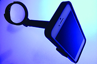
A team at Stanford University in California—Robert Chang, MD, Alexandre Jais, MS, and David Myung, MD, PhD—is developing a new inexpensive adapter dubbed “EyeGo,” which is designed to allow ophthalmologists and health-care providers to capture clear images of the eye using a smartphone and existing ophthalmic lenses. “Everyone is talking about adapters that hook your smartphone onto the slit lamp,” notes Dr. Chang, assistant professor of ophthalmology at Stanford University’s Byers Eye Institute. “We tried those, but they were cumbersome and took too long to set up. We wanted something you could just pull out of your pocket to capture an image of the eye instantly.
“Smartphone cameras have become more powerful, and ophthalmic lenses are very high quality, so we felt we could obtain an image of the retina by performing indirect ophthalmoscopy just using the phone and a 20-diopter lens,” he continues. “However, it was not easy to obtain a clear image of the eye just holding the two in midair. So we started experimenting with the simplest adapter possible to aid in aligning the phone and lens in the correct positions. We also knew portability was key.”
Dr. Chang says they’ve been using 3-D printers to create different adapter designs. “We’re constantly improving on the prototypes, and the 3-D printing component has helped us evolve faster,” he explains. “We’re trying to print every component.”
|
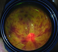
“Capturing photos of the back of the eye came next, which is a much more challenging goal—especially undilated images,” he continues. “We played around with the optics and lighting to achieve a dilated image of the retina, but we’re still working on undilated eyes.”
Working in the Field
After reaching this point, Dr. Chang says they began a couple of research studies. “The first was a proof of concept study—demonstrating that residents could take high-quality, useful photos in the emergency department or at the patient’s bedside in the hospital,” he explains. “In the clinic, we incorporated it into diabetic screening day, where we showed that the images were highly correlated with the indirect exam in determining which individuals needed a retina referral due to severe diabetic retinopathy.” Dr. Chang says more studies will be forthcoming.
The team also realized early on that they needed a secure and HIPAA-compliant way to label and organize the resulting photos. “We envisioned a telemedicine-type service for storage and interpretation,” he says. “This kind of service already exists for dermatology patients who can use a camera app to have a skin rash evaluated remotely. To accomplish this, we’re partnering with DigiSight Technologies in Portola Valley, Calif., another start-up with an app and secure cloud platform that helps providers share images and store and retrieve medical information.”
| ||||||
“Ultimately, it would be great if patients were able to take their own anterior photos using the EyeGo,” he adds. “Patients already send us smartphone selfie pictures of their eyes, but without an adapter the photo quality is usually not very good. Imagine every postop cataract patient being sent home with an adapter in case they experience a problem. The treating physician could better distinguish something routine from something serious. Of course, if people are concerned about their eye and they have access to a doctor, they’ll probably just go see the doctor. But someone in a rural area without easy access might be happy to have this alternative.”
Dr. Chang says they’d like to make the EyeGo adapters available as soon as possible, emphasizing that low price is a priority. “We’re currently working on the regulatory and liability issues, which hopefully will be solved in the coming months,” he says. “We want this to be available to everyone. We believe that’s how we can help the world the most. There are already a lot of smartphones out there, and if you could turn one into a medical triaging device capable of being used by anyone, that would be a remarkable breakthrough.”
A Low-cost Slit Lamp Adapter
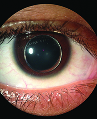
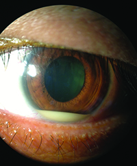
Another way to use smartphone photo technology, of course, is to capture the high-quality images produced by a slit lamp. In order to do that, several companies have created adapters designed to allow the surgeon to attach a smartphone directly to the oculars. However, most of these adapters range in price from $75 to more than $500. That inspired Jan Bond Chan, MBBS, who practices at Hospital Selayang and Hospital Universiti Sains in Malaysia, to create a simple alternative that can be easily assembled from inexpensive parts. (The total cost of creating his adapter is in the range of $15.)
“We tried using smartphones to take pictures directly through the eye piece of the slit lamp without an adapter, but getting a perfect picture was very difficult,” he explains. “Doing so usually required three people. The first person would hold the patient in place at the slit lamp and get him to keep his eye open. The second person would use both hands to stabilize the smartphone camera, and the third person would manage the zoom function and press the button to take the picture.” Dr. Chan realized an adapter was practical, but he wanted to create a simpler, less expensive alternative to those on the market.
| |||||||||
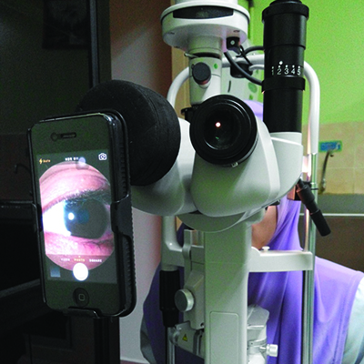
Dr. Chan’s adapter is constructed from four hard, cylindrical sponges that are about 1 cm thick, with a diameter at least 20 mm wider than the diameter of the slit lamp’s eyepiece, in conjunction with an inexpensive, removable smartphone case. (One online source for the 1-cm thick cylindrical hard sponges can be found at: daisojapan.com/p-20209-eva-cushion-d315-in-4-pcs-12pks.aspx.) Using the slit lamp’s eyepiece as a guide, circular holes are cut in three of the four sponges, which are stacked and glued together, forming a sponge tube that will slide over the eyepiece. A smaller hole is then cut in the fourth sponge; that hole will be centered over the smartphone camera lens. The fourth sponge is then added to the stack. The exact length of the resulting tube should match the focal length of your cameraphone.
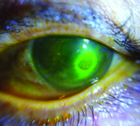
To complete the assembly, the sponge tube is slid onto on the functioning eyepiece. With the smartphone inside the case, the camera is carefully aligned to allow a clear, centered view through the adapter; the adapter is then glued to the smartphone case. A detailed description of the device and its assembly has already been published,1 and a YouTube video showing how the adapter is assembled can be seen at: youtube.com/watch?v=jwq7nDwgxa0&feature=youtu.be. (A picture of the adapter in use, and sample pictures taken using the adapter, can be seen above.)
“Initially I created the adapter for my own personal use,” says Dr. Chan. “However, my colleagues saw my invention and encouraged me to continue working on the design to improve it. I eventually entered a local innovation competition in Malaysia and won first prize for the design.”
Dr. Chan says one obvious disadvantage of his invention is that it may have to be recreated when a different phone is purchased. “The quality of picture you get also depends on the smartphone that you use,” he adds. “I do notice that the speed of the camera that captures the picture makes a big difference. The iPhones I’ve used will take a picture within half a second after I push the button. Some competing phones take as long as one second to capture the image. This makes a huge difference, as patients tend to move their eyes. Also, exposure and focus are very important to getting a great picture, so the built-in camera may need help from additional apps to get the best shot. For example, separating the focus area and the light exposure has required purchasing an additional app.”
Dr. Chan says he’s been using the adapter for about three years and has improved the design several times. “I use it about once a week,” he says. “Meanwhile, the first version of it is still being used and has yet to fall apart. I’ve introduced the adapter to four hospitals so far, and everyone is very happy with it.” REVIEW
The intellectual property for the EyeGo adapters is owned by the Stanford University Office of Technology and Licensing. For more information about EyeGo, please contact eyegotech@gmail.com.
1. Chan JB, Ho HC, Ngah NF, Hussein E. DIY Smartphone Slit Lamp Adapter. J Mobile Tech Med 2014;1:16-22.
