Performing penetrating keratoplasty in children poses a host of challenges that a corneal surgeon does not usually consider in an adult.
Our group at the
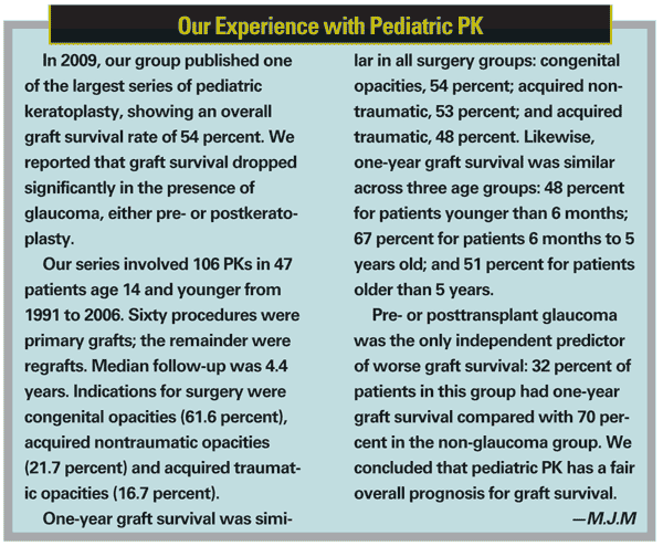
PK in children has become more commonly performed with better results, primarily because corneal surgeons have recognized the special surgical techniques and the potential problems specific to this population. The primary indications are congenital or acquired opacities, either traumatic or nontraumatic. However, the decision to perform PK depends not only on the presence or absence of corneal disease, but whether it is unilateral or bilateral as well as the age of onset as determining factors.
Because problems are significant in this undertaking, the surgeon must make difficult decisions at the outset about the likelihood of graft survival and whether the transplant will provide the child with functional improvement. The presence of glaucoma also factors significantly into decision making. Our study showed that patients with glaucoma had a one-year graft survival rate of 32 percent compared with 70 percent in patients without glaucoma. The overall one-year graft survival rate was 54 percent.
The family needs to understand the graft has about a 50-percent chance of survival. The risks in children are significantly different from adults, not only because of corneal clarity, but due to the potential for amblyopia.
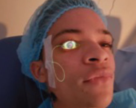
From the preoperative evaluation to the postoperative examinations and care, children with PK require a high level of care and attention. They must be placed under general anesthetic for the preoperative examination in order to obtain a detailed view of the back of the eye for an accurate assessment of the anterior segment and measurement of intraocular pressure, the latter of which is absolutely crucial in these patients.
Of course, the procedure itself demands the use of general anesthetic, and commonly the
postoperative procedures, including suture removal, may require the use of general anesthesia.
Surgical Technique
The pediatric eye differs vastly from the adult eye: It is considerably smaller; the sclera and cornea are more supple; and the anterior segment is more crowded. When incising the cornea, the globe in the child has a much greater tendency to collapse, the intraocular contents are more likely to come forward into the operative field. This makes both the removal of the old cornea and the attachment of the graft potentially more challenging than PK in the adult. To avoid these complications, we always use a scleral expansion ring such as a Flieringa ring to keep the eye intact during surgery.
Operating on the pediatric patient also demands a very slow and measured entry into the eye. We employ a technique to hinge the recipient cornea and place the donor cornea into position before removing the host cornea entirely. This provides a safety valve to cover the eye in case a problem arises between removal of the recipient and securing of the donor. The procedure requires very intense attention to detail between corneal removal and placement because of all that can happen in that interval.
A Team Approach
One of the take-home messages of our PK series is the essential role of the care team, comprising four components:
- a corneal surgeon experienced in pediatric grafting;
- involvement of a glaucoma consultant from the beginning;
- participation of a pediatric ophthalmologist from the beginning; and
- the full participation of the family.
The pediatric ophthalmologist participates in all postoperative management—refractive care, prescribing contact lenses and managing amblyopia, which results either from the initial process or the graft itself. Also, I cannot stress enough the importance of having the family on board. The pediatric PK recipient is a very high-maintenance patient, requiring frequent drop administration, management of patching and amblyopia treatment and monitoring, so that the graft is not traumatized.
The necessity for this team explains why so few corneal surgeons perform pediatric PK. Our results have been modest, but they were achieved under optimal conditions. When I go into the OR to do the examination under anesthesia, I bring along the glaucoma specialist and the pediatric ophthalmologist. If you do not have that team, you probably should not do pediatric PK.
Postoperative Care
The child's healing process is very different from the adult's. Suture removal in the child begins at six weeks postoperatively rather than six months. You can anticipate that the wound is going to heal much faster in the child, requiring earlier suture removal.
Contact lenses have a vital role in the postoperative management of pediatric PK patients. At least 50 percent of these patients, and perhaps more, ultimately require a contact lens for vision correction because the sutures induce corneal irregularity, which can induce amblyopia. Many of these patients also become aphakic during the procedure, which requires contact lens correction.
Future Considerations
While the technical approach for pediatric PK has remained essentially unchanged from 1991 when we began our published study, some innovations in the adult procedure that have not yet been wholly adopted for children are worth noting. A few cases of endothelial keratoplasty in infants with congenital endothelial dystrophy have been reported in the literature. This approach may, over time, become more common because it avoids many of the issues of the open eye in the pediatric graft patient.
Moreover, the femtosecond-laser assisted keratoplasty used in adults has not yet been tried in children. This approach may prove useful in the pediatric population, because it provides for a much stronger wound and earlier suture removal.
Based on our experience with PK in children, the key elements for success are a thorough understanding of the nuances of a child's physiology and behavior, a multidisciplinary team that fully involves the family and careful, methodical surgical technique.
Dr. Mannis is professor and chair of the Department of Ophthalmology & Vision Science at the
1. Huang C, O'Hara M, Mannis MJ. Primary pediatric keratoplastly: Indications and Outcomes. Cornea. 2009;28:1003-1008