When Chicago surgeon Lou Probst needed to document the apparent shift of flap position after using the IntraLase, he turned to off-the-shelf editing software, and then made some personal tweaks in order to get the results he wanted. Here’s how he put the software to use, and how you could do the same when documenting cases of your own.
A Mysterious Problem
Dr. Probst says he had been doing LASIK with the IntraLase for six years when he ran into Los Angeles surgeon Robert Maloney at a national ophthalmology meeting. Dr. Maloney informed him that he had begun noticing that his IntraLase flaps, though they looked centered under the IntraLase, were always temporally displaced when viewed under the CustomVue excimer laser. Dr. Maloney had even gone so far as to devise a nomogram to try to get them to appear to be more centrally positioned when the patient was under the excimer laser. This revelation came as a surprise to Dr. Probst, who had never noticed such a shift. Even so, he too began adjusting his flaps to try to compensate.
Dr. Probst later discussed the phenomenon with Long Island, N.Y., surgeon Eric Donnenfeld, who suggested that the apparent shift of the flap location might instead be a product of centroid shift of the pupil. “I thought that sounded like a reasonable explanation,” says Dr. Probst, who set out to test the theory. But to test it accurately, he had to go outside the realm of ophthalmology and into the realm of video and photo editing.
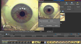 |
| Adobe Premier Elements allows surgeons to import surgical video and make screen captures of certain key parts to use for slides or to take measurements. |
Dr. Probst already had a DVD recorder (which costs about $200) attached to his operating microscope, so he had films of LASIK procedures. In order to capture parts of the video to manipulate in his computer, he purchased the video editing software Adobe Premiere Elements ($100). “The nice thing is that you put the DVD into your laptop, then go to Premiere’s ‘Get Media’ function, select ‘PC/DVD Drive,’ and it loads a section of video right there,” explains Dr. Probst. “I then use the program to capture particular still frames from the videos.” To measure pupil sizes and centroid shifts, he captured images of the eye under the wavefront aberrometer, then under the femtosecond laser, and finally under the excimer. “From there, I had to put the screen captures into Adobe Photoshop in order to make measurements,” he says.
Dr. Probst says using Photoshop was the challenging part, starting with getting the right version of the program in order to be able to make on-screen measurements. “I had the limited version, Photoshop Elements (which costs around $100), and looked for the ability to make measurements on the screen, but discovered it wasn’t in that version,” Dr. Probst recalls. To enable on-screen measurement, he needed to purchase the full version of the program, which is priced at about $500.
Dr. Probst was happy to finally be able to make measurements of the anatomical structures he was seeing on the screen, but was confounded by the fact that the program only allowed him to make measurements in pixels. This is when he had to get creative. “The trick was the measurement tool had to be calibrated,” he says. “So before I performed the procedure, I held a set of digital calipers, open to a set width, in front of the microscope and filmed them. Then in Photoshop, I could use the measurement tool to measure the calipers’ width in pixels, giving me a conversion factor of pixels to millimeters. I used that factor to measure all the different apparent shifts in flap position and pupil sizes in my surgical images.”
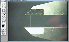 |
| To get a conversation factor to use for other measurements, Dr. Probst measured calipers in pixels in Photoshop and then converted the pixels to millimeters. |
Armed with still images from his surgeries and measurements of pupil width and centroid shifts for 10 eyes, Dr. Probst realized that the apparent decentration of the IntraLase flap was in fact an illusion caused by centroid shift of the pupil as it constricted under the increasing illumination of the various OR instruments. The flap was stationary. “The pupil was moving nasally and causing a perceived displacement in the flap,” he says. Specifically, the average pupil size decreased from 6.64 mm OD and 6.24 mm OS under the WaveScan aberrometer to
4.48 mm OD and 4.83 mm OS under the IntraLase and finally to 3.28 mm OD and 3.06 mm OS under the CustomVue’s bright illumination. Along with this constriction, the pupil centroid shift in the nasal direction in the x-axis was 0.35 mm OD and 0.20 mm OS.
“This was surprising to me, but then I realized that few surgeons notice that the pupil changes, and I didn’t initially detect the apparent shift of the flap position either,” says Dr. Probst. “So, after doing these measurements and imaging, my understanding of the procedure has gone full circle: from me not noticing it; to noticing it and adjusting my flaps; to realizing the flaps aren’t shifting at all. It’s an interesting example of how the pupil centroid shift affects our perception of what’s occurring with the created flap.”
The Next Step
Now that he’s got a useful digital editing and measurement scheme, Dr. Probst plans on using it for future studies, and says other surgeons can too. “One of the most interesting parts of all this is that I’m able to do it just using a laptop computer, not a standalone—possibly more powerful—desktop,” he says. “It has 4 GB of RAM, a 2.2-GHz processor and a 500-GB hard disk drive, and is running under Windows 7. Essentially any computer bought within the last year or two would be fine for this.”
He’s currently using the combination of Premiere and Photoshop to study oval flaps in LASIK. “With my next presentation, which I will give at the 2011 meeting of the American Academy of Ophthalmology, I’ll confirm that the cornea is, in fact, oval, rather than round, with an average ovality of 8 percent,” he explains. “Though we learned that the cornea is oval back in anatomy class, I think many of us come to think of it as round after we start practicing medicine. I then show measurements of the oval flaps created with the femtosecond and compare them to the oval flap settings on the AMO iFS system to show that the preop settings are indeed accurate; there’s no significant difference between what you measure and what you get.”
Though Dr. Probst says using Photoshop in years past seemed intimidating, and risked stranding the user “in a sea of information,” the later versions have become much more approachable. “I’ve found that the current versions of these programs are very user-friendly and aren’t difficult to understand,” he says.


