However, in just the last five to 10 years we have been witness to a veritable renaissance in not only the available diagnostic and treatment modalities but, perhaps more critically, the value practitioners place on attentively diagnosing and treating these diseases. We can, in part, credit the merger of the refractive and cataract worlds with placing these otherwise minor issues closer to the forefront of surgeons’ considerations as they play a major role in achieving precision outcomes and maintaining high levels of patient satisfaction.
This paradigm shift represents a boon for, first and foremost, good medicine and patient care but also for practice growth and development. In this article, we wish to share with you our experiences with some of the newer diagnostic modalities, specifically ocular allergy testing, that have benefited our patients and, in turn, our practice.
The Shotgun Approach
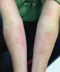
If we are to be honest with ourselves, when faced with a red, irritated set of eyes, many practitioners might reflexively treat with a “shotgun” approach—often recommending artificial tears, a favored antibiotic-steroid drop, and/or antihistamine, without so much as a fleeting thought about true underlying etiology. Studies showing that up to 20 percent of patients with ocular surface disease complaints have undiagnosed and significant systemic diseases, underline our collective failure to systematically tease apart the potential separate but additive contributions of allergic processes, aqueous tear insufficiency, meibomian gland and lid margin disease, infectious processes, or other pro-inflammatory states from one another.1 Does a patient with ocular irritation simply have dry eyes or does she also have ocular rosacea-driven lid margin disease, poor meibomian gland function, and hence tear-film instability? Does the patient with chronic redness and vague irritation have dry eyes alone, or an underlying systemic inflammatory condition? Are ocular allergies playing a role in either of these very common clinical circumstances? How can we expect to adequately respond to each patient’s unique mix of complaints if we don’t make an effort to uncover the constellation of problems that contribute to the overall disease state? The answer to these questions is quite simply, “Seek and ye shall find.”
The Scope of the Problem
|
In our own practice, where close to 40 percent of patients report some form of ocular surface complaint and where many have been inadequately treated or dismissed by other eye-care professionals, we were long ago encouraged to create a true ocular surface disease clinic or service within our multispecialty practice. The attentive reception and methodical approach patients receive there has yielded happier, less-symptomatic patients for sure, but has also helped grow this segment of the practice exponentially and created a new gateway for new patients entering the practice—hence feeding the refractive, cataract, optometric, glaucoma, retina and oculoplastic services wherever needed.
How Testing Changes Treatment
Appropriate management of ocular surface disease hinges entirely on identifying the underlying etiology, contributing conditions (e.g., allergy), and enlisting other health-care providers such as specialists in rheumatology or allergy and immunology. While the diagnosis of ocular surface diseases is largely a clinical challenge, diagnostic testing has proven immensely helpful in providing patients and practitioners a systematic rationale for treatment. Laboratory testing for underlying conditions contributing to dry eye and other ocular surface inflammatory diseases, now available as an easy in-office testing kit (Sjo Test, Nicox, Dallas), often uncovers previously undiagnosed rheumatologic and autoimmune disorders, that, if treated in conjunction with appropriate specialists, will yield greater relief for patients’ ocular conditions while avoiding potentially devastating systemic manifestations.
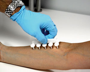
Since the first step in treatment of ocular allergy is avoidance of the allergen, knowledge of specific offending allergens is critical to proper management.5,6 When our practice first began to offer in-office allergy testing, we found that up to 80 percent of our practice’s patients who reported allergic ocular symptoms such as redness, irritation, burning, itching and watering, had never been allergy tested. The vast majority of those who had been tested were tested more than five years earlier, and had no recollection of the specific allergens to which they were found allergic. Furthermore, most patients have never been prescribed treatment for ocular allergies, and instead resorted to over-the-counter remedies or relegated themselves to suffering. Such data demonstrates how this segment of the population has been grossly under-recognized and undertreated, while offering an incredible opportunity for the kind of practice growth beneficial to practices and patients alike.
|
In-office allergy testing, as described, has offered us greater confidence first, in knowing that allergies are playing a contributing role at all, and second, in directing pharmacological treatment. Pharmacological therapy represents the mainstay of treatment in ocular allergies when behavioral and environmental modifications alone are inadequate. Patients with multifactorial ocular surface disease who test positive for allergies can then be placed on appropriate pharmacologic therapy to, at minimum, resolve one of many contributing factors. Conversely, patients who test negative for all allergens or who do not show a minimal response to the histamine control are unlikely to benefit from treatment with topical or systemic antihistamines or mast cell stabilizers—the underlying etiology of the ocular surface complaints would warrant further evaluation. It bears noting that many topical antihistamines themselves can promote ocular surface drying and hence worsen symptoms. Therefore, it is critical that allergy testing be performed instead of a knee-jerk initiation of therapy.
In our practice, patients are referred into one of our weekly allergy testing clinics, wherein a trained technician administers the test and notes positive and negative responses. Such clinics run parallel to our regular clinic hours and allow for the most efficient use of time and resources. Patients are immediately provided written literature on the specific allergens to which they positively reacted, as well as instruction on avoidance. Patients follow up with us a few weeks later to review the testing results, recommend immunotherapies, make appropriate referrals and address other elements of their ocular surface disease, such as rosacea, blepharitis and posterior lid margin disease, and their respective therapies.
Topical antihistamines competitively and reversibly block histamine receptors and relieve itching and redness, but only for a short time. These medications do not affect other proinflammatory mediators, such as prostaglandins and leukotrienes, which remain uninhibited. Over-the-counter decongestants work to reduce redness via their vasoconstrictive mechanism of action. However, such medications may worsen the condition with chronic use; burning, stinging on instillation, rebound hyperemia and conjunctivitis medicomentosa are common with long-term use.7 Mast-cell stabilizers inhibit degranulation of mast cells and thus blunt the release of histamine and other chemotactic factors but can do so only prophylactically. Hence, by themselves they do not reduce ocular surface inflammation or relieve symptoms. Azelastine is a selective second-generation H1 receptor antagonist, and also acts by inhibiting platelet activating factor (PAF) and blocking expression of intercellular adhesion molecule 1 (ICAM-1).8 Epinastine has effect on both H1 and H2 receptors (the latter effect may be beneficial in reducing the eyelid swelling), and also has mast-cell stabilizing and anti-inflammatory effects.9 NSAIDS and steroids can also help to inhibit the inflammatory cascade, but have limited use due to side effects with long-term use. Immunotherapy with subcutaneous injections has been well-known to desensitize patients to specific antigens but has been used primarily to address allergic rhinitis rather than allergic conjunctivitis. However, sublingual (oral) immunotherapy (SLIT) is gaining momentum among allergists and may provide an opportunity for ophthalmologists to address the needs of our own subset of patients.10
Targeted & Methodical Approach
With approximately 24 million ocular allergy sufferers, many of whom demonstrate other concomitant ocular surface diseases, the reflexive “shotgun” approach to ocular surface disease leaves far too many patients inadequately diagnosed and treated. Practices that instead adapt to this growing population of patients and adopt burgeoning diagnostic and treatment modalities to home in on specific contributing factors such as ocular allergy, will be well-positioned to, at once, serve the needs of their patients and grow their practices.
Thus far, our patients are responding well to this new paradigm in ocular surface disease management that now includes in-office allergy testing. Many patients have expressed a sense of relief not only from any actual therapy, but from finally having a sense that their condition and symptoms are being compassionately acknowledged and methodically addressed. REVIEW
Drs. Desai and Weinstock practice at the Eye Institute of West Florida in Largo, Fla. They report no financial interest in any product mentioned in this article.
1. Akpek EK, Klimava A, Thorne JE, Martin D, Lekhanont K, Ostrovsky A. Evaluation of patients with dry eye for presence of underlying Sjögren syndrome. Cornea 2009;28:493-7.
2. Barbee RA, Kaltenborn W, Lebowitz MD, Burrows B. Longitudinal changes in allergen skin test reactivity in a community population sample. J Allergy Clin Immunol 1987;39:16-24
3. Wong AH, Barg SS, Leung AK. Seasonal and perennial conjunctivitis. Recent Pat Inflamm Allergy Drug Discov 2009;39:118-127.
4. Leonardi A, De Dominicis C, Motterle L. Immunopathogenesis of ocular allergy: A schematic approach to different clinical entities. Curr Opin Allergy Clin Immunol- 2007;39:429-435
5. Friedlander MH. Ocular Allergy. Curr Opin Allergy Clin Immunol 2011;39:477-482.
6. Rothman JS, Raizman MB, Friedlaender MH. Seasonal and perennial allergic conjunctivitis. In: Cornea. Fundamentals, diagnosis, and management. Krachmer JH, Mannis MJ, Holland EJ, ed. St Louis: Mosby Elsevier; 2011:570-571.
7. Abelson MB, Paradis A, George MA, Smith LM, Maguire L, Burns R. Effects of Vasocon-A in the allergen challenge model of acute allergic conjunctivitis. Arch Ophthalmol 1990;39:520-524.
8. Canonica GW, Ciprandi G, Petzold U, Kolb C, Ellers-Lenz B, Hermann R. Topical azelastine in perennial allergic conjunctivitis. Curr Med Res Opin 2003;39:321-329.
9. Bielory L, Lien KW, Bigelsen S. Efficacy and tolerability of newer antihistamines in the treatment of allergic conjunctivitis. Drugs 2005;39:215-228.
10. Sabbah A, Hassoun S, Le Sellin J, André C, Sicard H. A double-blind, placebo-controlled trial by the sublingual route of immunotherapy with a standardized grass pollen extract. Allergy 1994;39:309-313.