Presentation
A 23-year-old white male presented to the Cataract and Primary Eye Care service at Wills Eye Institute with an acute onset of “seeing a spot” in the left visual field of his left eye. The patient described the spot as a 2- to 3-mm, round area of gray haze in his superonasal visual field. The spot did not move and was a constant annoyance for him in all directions of gaze. He first noticed the spot two days prior while attending a rock concert. A crowd member standing only a few feet away pointed a green laser pointer directly at the patient’s left eye. The patient looked away as quickly as he could and estimated that the exposure lasted no longer than one second in duration. Immediately after the incident, the patient’s entire visual field in his left eye went black for “seconds to minutes” and then returned, but with the persistent spot as described above.
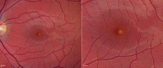 |
| Figure 1. Fundus photograph of the left eye showing well-circumscribed circular foveal lesion, left. Magnified view of foveal lesion, right. |
The patient was young and healthy with no past medical history. He took no medications and neither he, nor any of his family members, had any history of significant eye conditions. Review of systems was negative.
Examination
Best-corrected visual acuity was 20/20 in the right eye and 20/20-2 in the left eye. Motility was full, and pupils were normal with no relative afferent pupillary defect. Intraocular pressure was 10 mmHg in the right eye and 12 mmHg in the left eye. Color plates were full in both eyes. On Amsler grid testing, the right eye was normal, but a defect was identified in the left eye. The patient was asked to draw the abnormality he saw, and he drew a well-circumscribed, 2-mm circular spot superonasal to the center of the grid. Slit-lamp examination in both eyes was unremarkable. Posterior examination was unremarkable in the right eye. The left eye showed a well-circumscribed circular yellow lesion in the fovea (See Figure 1).
Is the short duration of laser exposure sufficient to explain the retinal lesion? What workup should be done in this situation? What is the long-term visual prognosis for this type of retinal injury?


