Presentation
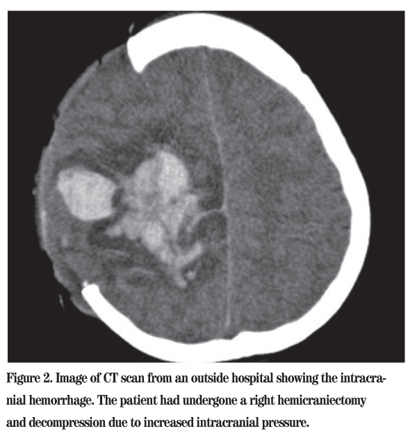
An otherwise healthy 14-year-old boy experienced pain in his right leg and a headache. Shortly after the headache began, he became unresponsive and collapsed. He was rushed to an outside hospital, where he was found to have an intracranial hemorrhage with increased intracranial pressure. He was taken directly to the operating room by the neurosurgical team, where he underwent a right hemicraniectomy and decompression. Postoperatively, he was arousable and responsive, and had residual left hemiparesis.
During his hospital stay, he noted decreased vision, difficulty focusing and also difficulty looking to the left with both eyes. He was examined by an outside ophthalmologist, who noted bilateral dense vitreous hemorrhage. The patient presented to Wills Eye Institute five months after the initial event. He still noted decreased vision OU and floaters OU. He did not see any flashes of light.
Medical History
Past medical history was unremarkable, and past ophthalmic history was significant only for high myopia.
Examination
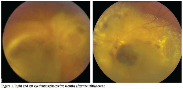
Visual acuity was count fingers at 1 foot OU, with no improvement to pinhole. His extraocular motility was full, there was no relative afferent papillary defect, and his pressures were 14 OD and 17 OS by applanation tonometry. His anterior exam was within normal limits OU. His anterior chamber was deep, and there was no inflammation. His dilated examination was as pictured (See Figure 1).
Diagnosis, Workup and Treatment
The differential diagnosis of vitreous hemorrhage in children includes blunt trauma, penetrating trauma and abusive head trauma. Neoplasm may also cause vitreous hemorrhage, as well as venous malformations, Terson's syndrome, inflammation such as pars planitis, regressed retinopathy of prematurity, X-linked retinoschisis and familial exudative vitreoretinopathy. The patient underwent several CT scans in the outside hospital (See Figure 2). A B-scan ultrasound examination was performed, whic h was highly suspicious for subretinal blood and fibrosis. This was later confirmed at vitrectomy and is believed to be the cause of the patient's subnormal visual acuity.
The patient was diagnosed with Terson's syndrome. The initial workup to determine the cause of the patient's intracranial hemorrhage, a rare spontaneous occurrence in someone at this young age, had already been performed. The differential diagnosis of intracranial hemorrhage includes trauma, blood dyscrasias, neoplasm, aneurysm, arteriovenous malformation, anemia, vitamin deficiency and hypertension. Lab studies revealed the patient to have a hemoglobin level of 7.8 and a platelet level of 2.0 x 10³ µL.
He was diagnosed with aplastic anemia. Despite a thorough workup, no definitive cause was found.
Discussion
Terson's syndrome was described by both Mortiz Litten in 1881 and Albert Terson in 1900. The diagnosis is made when a patient has both a subarachnoid hemorrhage and intraocular bleeding. It is often bilateral and usually secondary to the rupture of intracranial aneurysms. The intraocular hemorrhages may be preretinal, intraretinal and/or subretinal. This is not due to blood dissecting through the optic nerve sheath, but rather is believed to be caused by the transmission of increased intracranial pressure down through the intervaginal space of the optic nerve. The incidence of Terson's in patients with intracranial hemorrhage varies from 8 to 40 percent in different case series. It is believed to be lower in children. Those who do experience Terson's are more likely to have a history of sustained loss of consciousness, or coma, from their intracranial hemorrhage. These patients also have an increased mortality rate when compared to similar patients with intracranial hemorrhage but with normal ocular exams. The hemorrhage usually clears within one year. Patients may experience a number of complications, including cataract, retinal detachment, amblyopia, epiretinal membranes and other macular abnormalities.
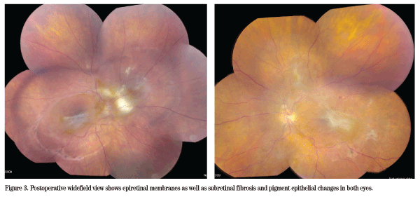
Some studies have shown no difference in visual outcomes with early vitrectomy compared to expectant management, but most studies do show improvement with early vitrectomy in patients with bilateral disease. Our patient underwent a pars plana vitrectomy several months after the initial event, and later underwent cataract extraction (See Figure 3). His current visual acuity is improved to 20/100 OD and 20/60 OS. He is back in school, and has since had his skull defect repaired.
The author would like to thank James F. Vander, MD, of the Wills Eye Institute Retina Service for his guidance with this case and manuscript.
1. Abu el-Asrar AM, al-Momen AK, Harakati MS. Terson's syndrome in a patient with acute promyelocytic leukemia on all-trans retinoic acid treatment. Doc Ophthalmol 1993;84(4):373-8.
2. Arroyo JG, Bula DV. Immunohistochemical Study of the Internal Limiting Mem-brane in Terson Syndrome. Retina 2004;24:155-157.
3. Aryan HE, Ghosheh FR, Jandial R, Levy ML. Retinal hemorrhage and pediatric brain injury: Etiology and review of the literature. J Clin Neurosci 2005;12:624-31.
4. Chan WM, Liu DT, Tham CC, Wu RM, Lam DS. Bilateral subhyaloid haemorrhage in aplastic anaemia. Br J Haematol 2003 Dec;123(5):757.
5. De Maeyer K, Van Ginderdeuren R, Postelmans L, Stalmans P, Van Calster J. Sub-inner limiting membrane haemorrhage: Causes and treatment with vitrectomy. Br J Ophthalmol 2007;91:850-2.
6. Kuhn F et al. Terson syndrome. Results of vitrectomy and the significance of vitreous hemorrhage in patients with subarachnoid hemorrhage. Ophthalmology 1998;105:472-7.
7.Lilley ER, Bruggers CS, Pollock SC. Papilledema in a patient with aplastic anemia. Arch Ophthalmol 1990;108:1674-5.
8. Mansour AM, Salti HI, Han DP, Khoury A, et al. Ocular Findings in Aplastic Anemia. Ophthalmologica 2000;214(6):399-402.
9. Margulies LF. Ocular manifestations of cardiovascular and hematologic disorders. Curr Opin Ophthalmol 1995 Dec;6(6):97-103.
10. Ritland JS, Syrdalen P, Eide N, Vatne HO, et al. Outcome of vitrectomy in patients with Terson's syndrome. Acta Ophthalmol Scand 2002 Apr;80(2):172-5.
11. Robbins and Cotran: Pathologic Basis of Disease, 7th ed.
12. Ryan SJ et al. Retina, 4th ed.
13. Spirn MJ, Lynn MJ, Hubbard GB 3rd. Vitreous Hemorrhage in Children. Ophthalmology 2006;113:848-52.
14. Shome D, Jain V, Jayadev C, Natarajan S. Scleral necrosis in a patient with aplastic anemia. Eye 2007 Jul;21(7):1017. Epub 2007 May 4.
15. Suzuki J, Goto H, Usui M, Sakai J. Serous retinal detachment in a patient with aplastic anemia associated with parvovirus B-19 infection. Graefe's Arch Clin Exp Ophthalmol. 2007;245:324-6.


