Every surgeon has his or her favorite tip or technique for getting the most out of LASIK and minimizing complications. Some have special approaches to the preop screening, while others make sure to handle the flap a certain way. Taken together, their tips can often help other surgeons get better results, too. Here are several surgeons' suggestions for better outcomes.
Before the Procedure
A big part of good results is acquiring a good wavefront image on which to base a custom procedure. Jonathan Davidorf, MD, of
Christopher Rapuano, co-director of
Though Dr. Davidorf prefers to perform a custom procedure on anyone who's a good candidate, he notes that it may not be necessary, and could be detrimental, in someone with very small pupils. "This is because most of the aberrations, particularly spherical aberration, are occurring at a wider diameter. If you do a wavefront treatment on an eye with a physiologic small pupil in low light, but you're basing your wavefront-guided treatment on a non-physiologic, dilated pupil, there could be some impact of that correction on the central zone. You'd hate to correct the unimportant higher-order aberrations that the patient never sees but potentially compromise the refractive outcome."
Cleveland Clinic's Director of refractive surgery Ron Krueger says that a wavefront map can be helpful even if you're not doing a custom treatment. "When putting in the patient's refractive numbers, I'd defintely use the wavefront map in terms of the amount and axis of astigmatism," he says. "That's been valuable. I've had scenarios where my manifest refraction, topography and cycloplegic refraction all had a different axis of astigmatism, but the wavefront helped me clinch where I should put the treatment."
Immediately before the procedure, Dr. Rapuano likes to explain to the patient, in detail, what he or she will experience during the procedure, so there are no surprises that might cause him to be uncomfortable or physically jerk during the LASIK. "I kind of shock him with a description of the microkeratome's buzzing by suddenly making a loud buzzing noise, so he'll be ready for the real thing," he says. To simulate the excimer ablation, he snaps his fingers loudly in the patient's face. "This really helps because patients know exactly what's going on," he says.
Intraoperative Ideas
Though the IntraLase laser has made flapmaking easier for many surgeons, there are ways to use it even more effectively.
"I've spent the last year trying to reduce or eliminate the opaque bubble layer that's associated with creating an IntraLase flap," says Louis Probst, MD, director of the
"So, the key is to get the meniscus small enough to create the flap, but big enough, at least at the area of the pocket, to touch the pocket," he continues. "You can do this through positioning, using applanation techniques such as soft docking and tilting the applanation cone a little so there's a little less pressure in the area of the pocket, leading to more meniscus in that area." (See "Get to Know Your Femtosecond Options" in this month"s issue for a discussion of soft docking.)
The other thing he's done is tinker with the laser settings to change the dimensions of the pocket. Since Dr. Probst is doing very thin, 100-µm flaps to get the benefits of the new procedure sub-Bowman's keratomileusis, he thought the pocket should reflect SBK's emphasis on being shallow rather than deep in the cornea. "Now, I create a pocket at 140 µm, rather than the standard depth of 225 µm," he says. "Also, the default size of the pocket is 0.25 mm, and I've increased it to 0.4 mm. With a larger pocket, it's much easier to have the meniscus touch it. Whenever you have that contact you're almost guaranteed to have no central OBL at all. It's been enough to really make a difference clinically, and my techs even asked me what I was doing differently because they noticed the absence of the OBL."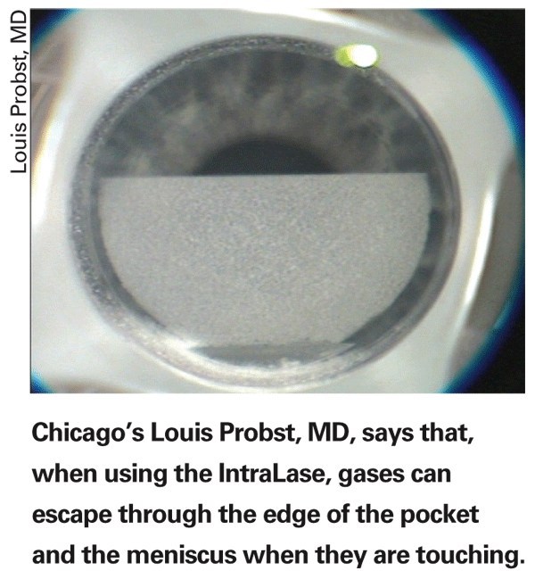
To help with centering the IntraLase, Stanford surgeon Edward Manche first moves the patient beneath the excimer microscope and marks the center of the entrance pupil with gentian violet. "When you place the suction ring, the eye can rotate," he explains. "And when you look down at it, because the eye's rotated you can get a parallax effect that makes it look like it's centered when it's not, and you get a decentered flap. So, if you mark the pupil center, then you'll know when it's on the eye that it's in the center of the alignment grid."
Manhattan
Postop Pearls
In the postop period, Dr. Krueger says it pays to be very careful when approaching enhancements, especially with the Alcon LADARVision system. "If I'm doing a custom retreatment, I'll definitely treat less than what the wavefront tells me," he says. "It's not uncommon to be faced with a custom retreatment of a patient who, though he previously had a high correction, now only has 0.75 D of myopia left and the computer tells you that there's 50 microns of tissue to be removed. By the Munnerlyn equation, even if your zone was 6.5 or 7 mm, this is about a 4-D correction! For these situations, you need to use as much offset as you can to change the target refraction. The LADAR system allows up to a 2.5-D offset."
Dr. Krueger is making 100- or even 90-µm flaps, and says he's starting to approach epithelial defects differently in these thin flaps. "If you have a little epithelial defect in your flap at the end of surgery, there's a good likelihood you're going to have some diffuse lamellar keratitis in the early postop period, because the cells are coming through the defect and migrating into the interface," he explains. "And if you have some DLK or epithelial ingrowth, you have to be quicker to lift the flap and irrigate than if you were using a thicker flap. This is because if you begin to get some lysis of the corneal tissue from the enzymes and inflammatory cells that are present, this action has a tendency to erode much more quickly through this thin flap than a thicker one."