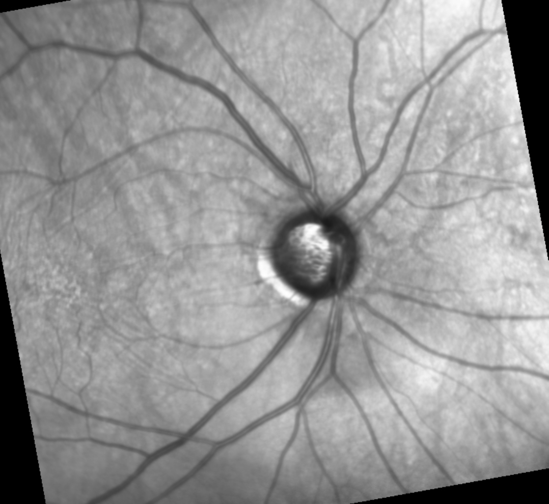
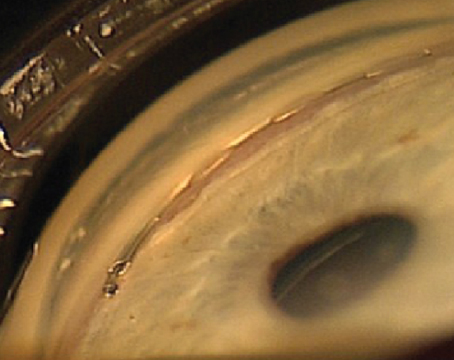
Nearly 30 to 40 percent of all normal adults, and almost all primary open-angle glaucoma patients are steroid responders,1 demonstrating clinically significant elevated intraocular pressures following corticosteroid use. It’s important to intervene early in the course of steroid-induced glaucoma to prevent irreversible optic neuropathy and vision loss. This condition can be tricky to treat in patients who require continued steroid therapy for underlying conditions.
Though it’s considered a form of open-angle glaucoma, steroid-induced glaucoma has a different pathophysiology from primary open-angle glaucoma.
Here, I’ll discuss why these two diseases may require different management approaches.
Risk Factors for a Steroid Response
Key risk factors for a steroid response include high myopia, type 1 diabetes mellitus, connective tissue disorders (e.g., rheumatoid arthritis), pigment dispersion, traumatic angle recession,2 primary open-angle glaucoma, prior penetrating keratoplasty, the duration of steroid therapy and the steroid’s anti-inflammatory potency.
Age is also a risk factor. Children have a strong intraocular pressure response to topical steroids,3 and younger patients (under 10 years old) are at the greatest risk for steroid-induced glaucoma. This glaucoma subtype accounts for about a quarter of all acquired glaucoma in children.4
 |
| The risk for steroid-induced glaucoma is partially influenced by the site of administration. In general, the closer to the center of the eye, the greater the risk. The steroid’s anti-inflammatory potency and the duration of steroid therapy also play a role. |
Route of Administration
As a general rule of thumb, the closer to the center of the eye the steroid is administered, the greater the risk. So, for example, steroid response risk is very low with intra-articular joint and intranasal administration and increases with inhaled, oral, intravenous, topical, periocular and intraocular administration.
- A meta-analysis on intranasal corticosteroids (484 studies with 10 RCT) reported a relative risk of 2.24 (95% CI, 0.68 to 7.34 percent) compared with placebo.5 The absolute increase in incidence of elevated intraocular pressure was 0.8 percent (95% CI, 0 to 1.6 percent) compared with placebo in non-glaucomatous individuals—a risk of less than 1 percent.
- A single case report described elevated intraocular pressure with inhaled steroids in a young child.6
- The risk with systemic administration of steroids varies, but it’s been reported to be low, ranging from less than 1 percent to 10 percent.7
- About 3 to 4 percent of patients treated with topical difluprednate experience a significant IOP increase >10 mmHg above baseline to 21 mmHg or more.8-9 Steroid-induced IOP responses after corneal transplantation are also well documented.10-11 Steroids are readily absorbed by the thin skin of the eyelids and around the eyes—anecdotally, many ophthalmologists have heard of a physician self-treating with a steroid face cream who comes in with pressures of 40 mmHg and massive vision loss.
- Periocular administration carries a significantly greater risk of steroid response compared with topical steroids.12 The sub-Tenon’s route has a significantly high risk, likely due to its proximity to the anterior chamber angle.13 A multicenter study of individuals treated with cortisone for uveitis demonstrated a 35-percent incidence of IOP elevation >24 mmHg.14
- For intravitreal administration, there’s a reported 12- to 15-percent risk of a >10-mmHg IOP rise after Ozurdex (dexamethasone), and an up to 50-percent risk with intravitreal triamcinolone acetonide.15
Two Unique Pathophysiologies
Though they share a mechanism of increased aqueous outflow resistance, steroid-induced glaucoma and primary open-angle glaucoma aren’t the same disease. Unlike primary open-angle glaucoma, steroid-induced glaucoma typically resolves spontaneously once steroids are discontinued. Based on their respective pathophysiologies, different approaches to treatment may be warranted.
• Primary open-angle glaucoma. The mechanism of primary open-angle glaucoma involves an increase of transforming growth factor beta-2 (TGFB2), which causes dysregulation of the extracellular matrix within the juxtacanalicular trabecular meshwork.
Another probable mechanism of this glaucoma subtype may be decreased trabecular meshwork cellularity.16 However, this mechanism has been observed only in cadaver eyes that were medically treated and/or had previously undergone surgery, not in untreated trabecular meshwork tissue. So, it’s unclear whether decreased cellularity was the result of the disease itself or its treatment. Another challenge with this theory is that the rate of cellularity decline is the same for non-glaucomatous eyes. If decreased cellularity were disease pathophysiology, one would expect the rate of cell loss to be greater in glaucomatous eyes.
• Steroid-induced glaucoma. One way steroids increase resistance to outflow by inducing dysregulation of the actin cytoskeleton (cross-linked actin networks [CLANs]). CLANs cause endothelial cellular stiffness and subsequently trigger an IOP increase.17 This has been observed within trabecular meshwork cells in cell cultures, cadaveric organ perfusion and in live mice. Very limited evidence suggests CLAN formation may also play a role in primary open-angle glaucoma pathophysiology, but the mechanism hasn’t been observed in untreated trabecular meshwork tissue.
In addition to CLAN formation, steroid-induced glaucoma involves dysregulation of the extracellular matrix, but this dysregulation is markedly different from that of primary open-angle glaucoma when seen on electron microscopy. Steroid-induced glaucoma exhibits more extracellular matrix accumulation in the juxtacanalicular region, as well as increases in curly collagen 6 and inhibition of matrix metalloproteinase enzymes from increasing levels of TIMPs. Additionally, a buildup of fine fibrillar material resembling fingerprints occurs, along with collagen IV, heparin sulfate, fibronectin, and an increase in other matrix proteins such as decorin, myocilin, fibrillin and secreted frizzle-related protein.
Medical Management
Discontinue steroids first, if possible. If the patient has been on steroid therapy for more than 18 months, IOP elevation may last for several weeks.18 Topical steroids can be switched to a lower potency. Then, start a prostaglandin analog—either topical or sustained release bimatoprost.
I recommend trying a rho kinase inhibitor sooner, rather than following the usual stepping pattern after a prostaglandin analog to the medications that decrease aqueous, because the rho kinase inhibitor mechanism of action potentially targets the pathophysiology of steroid-induced glaucoma.
Rho kinase inhibitors affect the actin cytoskeleton by disrupting the actin stress fibers and focal adhesions in trabecular meshwork cells.19-20 In a steroid-induced OHT mouse model study published earlier this year (n=56), morphological changes (extracellular matrix accumulation) and reduced trabecular meshwork effective filtration area induced by dexamethasone were partially reversed after one week of treatment with a rho kinase inhibitor (p<0.05) or five-week discontinuation of dexamethasone (p<0.01).21 This correlated with reduced IOP. IOP reductions were greater in rho kinase inhibitor-treated eyes than eyes that discontinued dexamethasone treatment.
Prostaglandin analogs should remain the first-line treatment, however, because they impact MMPs and TIMPs, targeting another part of steroid-induced glaucoma’s pathophysiology. These drugs shift the MMP/TIMP balance in the ciliary body and the trabecular meshwork toward greater turnover of extracellular matrix.22-29
Selective Laser Trabeculoplasty
It’s unclear whether there are any meaningful differences between primary open-angle glaucoma and steroid-induced glaucoma with regard to SLT success. A retrospective study reported a 72-percent SLT success rate (≥20 percent reduction from baseline IOP) in 25 eyes with steroid-induced glaucoma.30
Another retrospective study (n=608 eyes) reported that steroid-induced glaucoma patients did better with SLT than primary open-angle (p=0.005) or pseudoexfoliation (p=0.01) glaucoma patients, but a significantly higher baseline IOP for steroid-induced glaucoma eyes may have been a confounder.31
In the same study, the two-year failure rate for steroid-induced glaucoma patients was 54-percent failure, compared with 84 percent (p=0.01) for pseudoexfoliation glaucoma and 84 percent for primary open-angle glaucoma (p=0.005).
Surgical Management
There’s a paucity of literature specifically comparing the efficacy of our incisional surgical procedures in POAG versus steroid-induced glaucoma.
• Goniotomy. A retrospective study of steroid-induced glaucoma versus primary open-angle glaucoma using Trabectome found no difference in the survival curve.32 A greater IOP response was seen in the steroid-induced eyes, but they also had a much higher baseline IOP—a problem with retrospective cohort comparisons.
The incidence of steroid response after either Trabectome or iStent goniotomy is about 12 percent.33 Greater axial length, low-tension glaucoma and traumatic glaucoma were risk factors for steroid response after goniotomy. In eyes with axial length >25 mm, the incidence was 40 percent.
• GATT. A retrospective chart review of 13 patients demonstrated that GATT was effective for steroid-induced glaucoma,34 which isn’t surprising. GATT resulted in a significant IOP reduction at all postop visits, with all patients experiencing IOP reduction >20 percent at two years. The number of glaucoma medications also decreased significantly from an average of 3.1 medications preoperatively to 0.8 medications at the last follow-up.
Another retrospective chart review of 46 eyes reported that GATT was effective for short-duration steroid-induced glaucoma.35 IOP decreased from mean 30.8 ±8.3 mmHg at baseline to 11.2 ±2.6 mmHg after one to two years (n=28). At the final follow-up, 45 eyes had IOP <21 mmHg and 39 eyes had IOP <18 mmHg with or without medication. Steroid response wasn’t seen in any eyes after surgery. A few other case reports have reported GATT efficacy.36-37
In conclusion, SLT and incisional procedures have comparable efficacies in steroid-induced glaucoma and primary open-angle glaucoma. Anecdotally, I’ve observed that trabeculectomies do better if the depot steroid remains in the eye. For medical management, I recommend using a prostaglandin analog as a first-line agent because these drugs alter the MMP/TIMP balance and can reduce the buildup of extracellular matrix. Consider using a rho kinase inhibitor as a second-line agent after the prostaglandin analog because this drug class directly affects CLANs.
Dr. Rhee is a professor and chair of the Department of Ophthalmology and Visual Sciences at Case Western Reserve University School of Medicine. He is also the director of the Eye Institute, University Hospitals of Cleveland. He receives researching funding from Allergan/Abbvie, Glaukos, Ivantis/Alcon, Cleveland Eye Bank Foundation and Ocular Therapeutix. He is also an ad hoc consultant for Alcon, Allergan, Avellino and Iantrek.
Dr. Singh is a professor of ophthalmology and chief of the Glaucoma Division at Stanford University School of Medicine. He is a consultant to Alcon, Allergan, Santen, Sight Sciences, Glaukos and Ivantis. Dr. Netland is Vernah Scott Moyston Professor and Chair at the University of Virginia in Charlottesville.
1. Dibas A Yorio T. Glucocorticoid therapy and ocular hypertension. Eur J Pharmacol 2016:787;57-71.
2. Jones III R, Rhee DJ. Corticosteroid-induced ocular hypertension and glaucoma: A brief review and update of the literature. Curr Opin Ophthalmol 2006;17-163.
3. Ohji M, Kinoshita S, Ohmi E, et al. Marked intraocular pressure to instillation of corticosteroids in children. Am J Ophthalmol 1991;112:450–454.
4. Kaur S, Dhiman I, Kaushik S, Raj S, Pandav SS. Outcome of ocular steroid hypertensive response in children. J Glaucoma 2016;25:4:343-347.
5. Valenzuela CV, et al. Intranasal corticosteroids do not lead to ocular changes: A systematic review and meta-analysis. Laryngoscope 2019:129;6-12.
6. Desnoeck M, Casteels I, Casteels K. Intraocular pressure elevation in a child due to the use of inhalation steroids—A case report. Bull Soc Belge Ophtalmol 2001;280:97–100.
7. Ritch R, Shields MB, Krupin T. The Glaucomas: Basic Sciences, 2nd ed. Mosby-Year. 1996.
8. Korenfeld MS, Silverstein SM, Cooke DL, Vogel R, Crockett RS; Difluprednate ophthalmic emulsion 0.05% (Durezol) Study Group. Difluprednate ophthalmic emulsion 0.05% for postoperative inflammation and pain. J Cataract Refract Surg. 2009;35:1:26-34.
9. Smith S, Lorenz D, Peace J, McLeod K, Crockett RS, Vogel R. Difluprednate ophthalmic emulsion 0.05% (Durezol) administered two times daily for managing ocular inflammation and pain following cataract surgery. Clin Ophthalmol 2010;4:983-991.
10. Kornmann HL, Gedde SJ. Glaucoma management after corneal transplantation surgeries. Curr Opin Ophthalmol 2016;27:2:132-139.
11. Price MO, Price DA, Price FW Jr. Long-term risk of steroid-induced ocular hypertension/glaucoma with topical prednisolone acetate 1% after Descemet stripping endothelial keratoplasty. Cornea 2023. Published ahead of print, June 7.
12. Chew EY, Glassman AR, Beck RW, et al. Ocular side effects associated with peribulbar injections of triamcinolone acetonide for diabetic macular edema. Retina 2011;31:2:284-289.
13. McGhee CN, Dean S, Danesh-Meyer H. Locally administered ocular corticosteroids: benefits and risks. Drug Saf 2002;25:1:33-55.
14. Sen HN, Vitale S, Gangaputra SS, Nussenblatt RB, Liesegang TL, Levy-Clarke GA, Rosenbaum JT, Suhler EB, Thorne JE, Foster CS, Jabs DA, Kempen JH. Periocular corticosteroid injections in uveitis: Effects and complications. Ophthalmology 2014;121:11:2275-86.
15. Rhee DJ, Peck RE, Belmont J, et al. Intraocular pressure alterations following intravitreal triamcinolone acetonide. Br J Ophthalmol 2006;90:999-1003.
16. Alvarado J, Murphy C, Juster R. Trabecular meshwork cellularity in primary open-angle glaucoma and nonglaucomatous normal. Ophthalmology 1984;91;564.
17. Rohen JW. Why is intraocular pressure elevated in chronic simple glaucoma? Ophthalmology 1983;90:7:758-765.
18. Sihota R, Konkal VL, Dada T, Agarwal HC, and Singh R. Prospective, long-term evaluation of steroid-induced glaucoma. Eye 2008;22:1:26–30.
19. Lin CW, Sherman B, Moore LA, et al. Discovery and preclinical development of netarsudil, a novel ocular hypotensive agent for the treatment of glaucoma. J Ocul Pharmacol Ther 2018;34;1-2:40-51.
20. Keller KE, Kopzcynski C. Effects of netarsudil on actin-driven cellular functions in normal and glaucomatous trabecular meshwork cells: A live imaging study. J Clin Med 2020 Oct 31;9:3524.
21. Ruiyi R, Humphrey AA, Kopczynski C, Gong H. Rho kinase inhibitor AR-12286 reverses steroid-induced changes in intraocular pressure, effective filtration areas and morphology in mouse eyes. Invest Ophthalmol Vis Sci 2023;64:7.
22. Weinreb RN, Kashiwagi K, Kashiwagi F, Tsukahara S, Lindsey JD. Prostaglandins increase matrix metalloproteinase release from human ciliary smooth muscle cells. Invest Ophthalmol Vis Sci 1997;38:2778-2780.
23. Anthony TL, Lindsey JD, WeinrebRN. Latanoprost’s effects on TIMP-1 and TIMP-2 expression in human ciliary muscle cells. Invest Ophthalmol Vis Sci 2002;43:3705-3711.
24. Weinreb RN, Lindsey JD. Metalloproteinase gene transcription in human ciliary muscle cells with latanoprost. Invest Ophthalmol Vis Sci 2002;43;716-722.
25. Oh DJ, Martin JL, Williams AJ, Peck RE, Pokorny C, Russell P, Birk DE, Rhee DJ. Analysis of expression of matrix metalloproteinases and tissue inhibitors of metalloproteinases in human ciliary body after latanoprost. Invest Ophthalmol Vis Sci 2006;47:953-963.
26. Oh DJ, Martin JL, Williams AJ, Russell P, Birk DE, Rhee DJ. Effect of latanoprost on the expression of matrix metalloproteinases and their tissue inhibitors in human trabecular meshwork cells. Invest Ophthalmol Vis Sci 2006;47:3887-3895.
27. Ooi YH, Oh DJ, Rhee DJ. Effect of bimatroprost, latanoprost, and unoprostone on matrix metalloproteinases and their inhibitors in human ciliary body smooth muscle cells. Invest Ophthalmol Vis Sci 2009;50:5259-5265.
28. Heo JY, Ooi YH, Rhee DJ. Effect of prostaglandin analogs: Latanoprost, bimatoprost, and unoprostone on matrix metalloproteinases and their inhibitors in human trabecular meshwork endothelial cells. Exp Eye Res 2020;194:108019.
29. Stamer WD, Perkumas KM, Kang MH, Dibas M, Robinson MR, Rhee DJ. Proposed mechanism of long-term intraocular pressure lowering with the bimatoprost implant. Invest Ophthalmol Vis Sci 2023;64:15.
30. Al Obaida I, Al Owaifeer AM, Aljasim L, et al. Outcomes of selective laser trabeculoplasty in corticosteroid-induced ocular hypertension and glaucoma. Euro J Ophthalmol 2022;32:3:1525-1529.
31. Zhou Y, Pruet CM, Fang C, Khanna CL. Selective laser trabeculoplasty in steroid-induced and uveitis glaucoma. Can J Ophthalmol 2022;57:4:277-283.
32. Dang Y, Kaplowitz K, Parikh HA, et al. Steroid-induced glaucoma treated with trabecular ablation in a matched comparison with primary open-angle glaucoma. Clin Exp Ophthalmol 2016;44:783-788.
33. Abtahi M, Rudnisky CJ, Nazarali S and Damji KF. Incidence of steroid response in microinvasive glaucoma surgery with trabecular microbypass stent and ab interno trabeculectomy. Can J Ophthalmol 2022;57:3:167-174.
34. Boese EA, Shah M. Gonioscopy-assisted transluminal trabeculotomy (GATT) is an effective procedure for steroid-induced glaucoma. J Glaucoma 2019;28:9:803-807.
35. van Rijn LJR, Eggink CA, Janssen SF. Circumferential (360°) trabeculotomy for steroid-induced glaucoma in adults. Graefes Arch Clin Exp Ophthalmol 2023;261:7:1987-1994.
36. Nazarali S, Cote SL, Gooi P. Gonioscopy-assisted transluminal trabeculotomy (GATT) in postpenetrating keratoplasty steroid-induced glaucoma: A case report. J Glaucoma 2018;27:10:e162-e164.
37. Hopen ML, Gallardo MJ, Grover DS. Gonioscopy-assisted transluminal trabeculotomy in a pediatric patient with steroid-induced glaucoma. J Glaucoma 2019;28:10:e156-e158.


