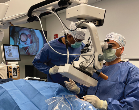
No matter the surgery, there’s always the potential for complications, and keratorefractive procedures—such as PRK, LASIK, and SMILE—are no exception. Ongoing advances in technology have improved the safety of these surgical approaches and lessened the risk of complications; however, surgeons must still be prepared to manage any issues that arise.
Below, we’ll delve into various complications associated with these procedures and discuss how to manage patients who undergo keratorefractive surgery while optimizing visual outcomes.
Intraoperative Complications
There are a number of complications—both intraoperative and postoperative—associated with LASIK and PRK. Most of the possible intraoperative complications of keratorefractive surgeries, according to Rockville, Maryland, cornea/refractive surgeon Okezie Igboeli, MD, occur during flap creation.
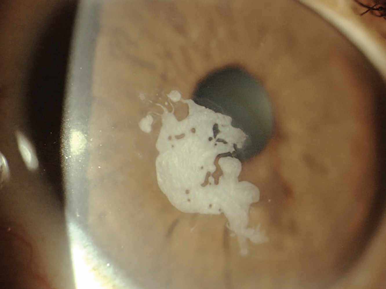 |
| Epithelial ingrowth in the visual axis needs an escalated response, say surgeons. |
• Flap complications. While the development of new technologies and techniques have made flap-related issues less common, these complications can still occur. Possible intraoperative complications include irregular, incomplete, decentered or buttonhole flaps.
Incomplete flaps can happen for a variety of reasons, such as a malfunctioning flap-making device or suction loss. “With some laser platforms, the surgeon may be able to re-dock and restart/continue the flap creation,” says Dr. Igboeli. “Otherwise, the cornea is allowed to heal, and LASIK or surface ablation may be attempted at a later date. If suction is lost during the side cut, the surgeon may be able to re-do the side cut only, or manually complete the side cut if suction loss occurred during the last seconds of the side cut.”
In the case of a decentered flap, which can be caused by a misaligned suction ring, surgeons should turn off suction and the ring should be repositioned. If there are repeated attempts without success, waiting five to 10 minutes can allow the “decentered gutter-like impression to disappear.”1
When there’s an interruption of the passage of the microkeratome across the cornea, buttonhole flaps can occur. “This can result from poor suction, problems with the blade, [and] steep corneal curvatures,” says Dr. Igboeli. “If a buttonhole is noted in the flap, the flap shouldn’t be lifted. However, if the flap is lifted prior to noticing the defect, the flap should be replaced, and bandage contact lens should be applied to the eye.”
Although rare, flap tears are another potential complication that can happen during the flap lift. This is more commonly observed in femtosecond LASIK procedures. Risk factors include a large diameter flap with corneal pannus, re-treatment procedures, the presence of a corneal scar and faulty instruments.1
Surgeons note that flap tears at the hinge can result in a free cap. In case of a small peripheral tear, the flap should be dissected away from the tear, recommends one study of complications. However, the authors go on to add, if it involves the visual axis, the procedure should be aborted and followed by re-treatment with surface ablation.1
• Free cap. In rare instances, a free cap can be created during LASIK. This can occur, according to Dr. Igboeli, when a microkeratome doesn’t stop at the predetermined point. Risk factors include flat corneas (less than 40 D) or improperly assembled microkeratomes.
“In such instances, if the stromal bed is regular and large enough for the laser treatment, the treatment can be completed,” notes Dr. Igboeli. “The free cap should be kept in a moist chamber while the treatment is taking place. The flap is then replaced ensuring that the epithelial side is up, and a bandage contact lens should be placed over the flap.
“If, however, the free cap is a smaller size without a large enough stromal bed, the free cap is replaced and a BCL placed,” he explains. “Surface ablation can be attempted at a later date (usually at least three months) after the cornea has healed.”
• Vertical gas breakthrough. In some cases, the gas released during flap creation with the femtosecond laser essentially creates a buttonhole, explains Dr. Igboeli. “Ideally, when this happens, the flap shouldn’t be lifted,” he says. “Just as with buttonholes, if the flap is lifted before the defect is noticed, the flap should be replaced and a BCL placed over the cornea. After the defect has healed, surface ablation may be attempted.”
There are rare instances when the gas released during flap creation can travel into the anterior chamber. If large enough, the bubbles can interfere with the tracking system of the laser, Dr. Igboeli notes. As a result, surgeons may have to wait a few hours for the bubbles to decrease in size before continuing the procedure. Placing ice packs over the affected eye can help speed up this process.
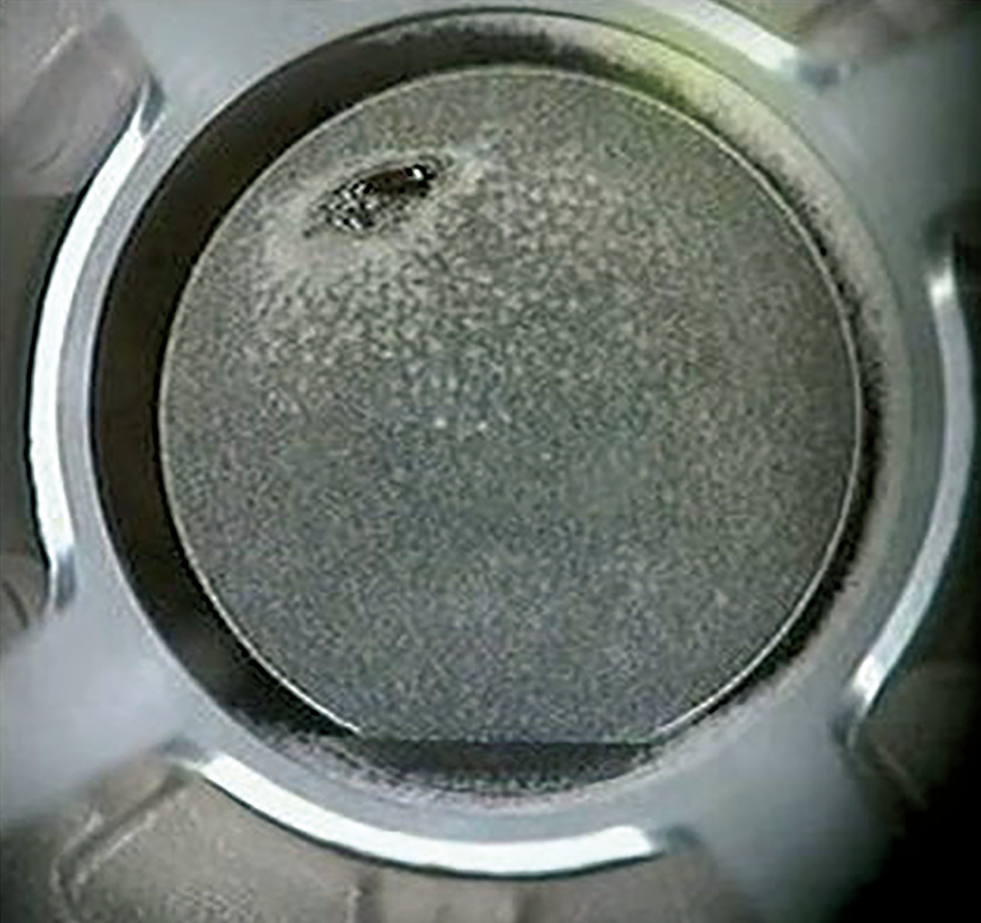 |
| A corneal abnormality can cause vertical gas breakthrough with the femtosecond. |
• Corneal perforation. This rare yet devastating LASIK complication can occur during flap creation. This can be the result of a microkeratome that’s not properly assembled, which was a requirement in older models. In older microkeratomes, improper placement of the depth plate could lead to perforations. “If using a microkeratome, it’s extremely important to ensure that it is properly assembled prior to use,” emphasizes Dr. Igboeli. “Newer microkeratomes come with fixed depth plates to avoid this complication.”
When corneal perforation occurs, surgeons say that suction should be immediately stopped.1 Conservative management by repositioning the flap and placing a BCL is done for small perforation; however, a large perforation requires surgical repair under sterile conditions.1
Postoperative Complications
Following PRK and LASIK, careful management and postoperative care is critical to address potential complications.
• Over- and undercorrections. Whenever the refractive target isn’t hit, it’s important to review all the preoperative data to ensure that the intended treatment was performed and that it was the correct treatment for the patient, notes Dr. Igboeli, who encourages surgeons to resist the urge to rush to an enhancement. “Wait at least three to six months before moving forward with enhancements, and always rule out ectasia prior to performing enhancement procedures,” he recommends. “In older patients, consider the possibility that the change in refractive error may be from progression of cataracts.”
• Corneal haze. While the risk of this PRK complication has lessened, corneal haze is still an issue that can present in clinical practice. Therefore, comprehensive management is critical and that begins with a work-up with imaging, including anterior OCT with two views of the corneal cross section, 9-mm epithelial map and corneal densitometry using Scheimpflug technology, according to Majid Moshirfar, MD, a professor of ophthalmology at the John A. Moran Eye Center, University of Utah School of Medicine, and research medical director at Hoopes Vision Research Center.2
Determining the best treatment approach will depend on several factors, including whether or not a patient presents with early- or late-onset haze. “Early-onset haze—within the first six months—may respond better to intensive steroid therapy, while late-onset haze may require a shorter steroid regimen or surgical intervention,” says Dr. Moshirfar, who is also the medical co-director of the Utah Lions Eye Bank. “Surgical options include mechanical debridement or superficial phototherapeutic keratectomy for superficial haze, and deep PTK or therapeutic myopic PRK ablation for deeper haze.”
Ophthalmologists should also consider preventive measures, such as intraoperative mitomycin C applied immediately after the PRK ablation. In 2021, researchers conducted a meta-analysis to analyze surgical outcomes in cases in which MMC was used intraoperatively after PRK for myopia and myopic astigmatism.3 They looked at 2,232 eyes that were prophylactically treated with MMC and 1,304 control eyes. The investigators found that MMC decreased the rate of both early and late onset corneal haze, with less loss of VA postoperatively in the treated group.
The duration of MMC application may vary based on ablation depth, according to Dr. Moshirfar, who is currently studying MMC’s effect on post-PRK haze.2
• Dry eyes. A common adverse event of laser refractive surgery, most patients will return to their baseline level of dryness by three to six months after the procedure, according to Dr. Igboeli. Screening for dry eyes or signs of ocular surface disease is important prior to surgery, however, note surgeons. Additionally, patient education and optimization of the ocular surface pre- and post-procedure is a priority. This includes maximizing tear film stability prior to the procedure. Effective management involves assessment of the eyelid, tear-film breakup time, rose bengal staining, corneal esthesiometry and Schirmer test. There are a number of treatment options, which have been covered extensively in various publications.4
• Decentered ablations. Optimal visual outcomes following LASIK and PRK depend on a number of factors, including good centration during excimer treatment, notes Dr. Igboeli.
“A decentered ablation can occur with improper positioning of the patient’s head, or with drifting of the patient’s eye during treatment,” he says. “Modern tracking systems have reduced the risk and incidents of decentration; however, it’s important to take note of conditions that can lead to decentration such as high refractive correction because of the longer time that it takes for treatment, and hyperopic ablations.”
When decentered ablations occur, topography-guided technology can help correct the potential irregular astigmatism, suggests Dr. Igboeli.5 RGP contact lenses and miotics for the reduction of optical aberrations from a decentration are management options surgeons can consider when addressing this complication.1
• Diffuse lamellar keratitis. An acute sterile inflammatory response following LASIK, DLK generally responds to topical corticosteroid treatment; however, when left unchecked, or in severe cases, there can be flap melting or severe scarring, explains Dr. Igboeli.
DLK is classified into four stages based on disease progression, this condition requires early recognition and prompt treatment. The stages, as defined by the American Academy of Ophthalmology:
— Stage 1 typically arises one to two days after refractive surgery. It’s characterized by peripheral inflammatory infiltrates without central corneal involvement.
— Stage 2 typically arises on postoperative days three or four, when inflammatory cells begin migrating from the periphery into the central cornea often compromising visual acuity.
— Stage 3 is characterized by further centripetal migration of inflammatory cells and the development of permanent corneal scarring. Stage 3 is often referred to as the “threshold” DLK subtype because of the likelihood that eyes in this stage will develop permanent scarring and a resultant loss of visual acuity.
— Stage 4 describes the phase in which stromal melting and further corneal scarring occur. The significant epithelial destruction that ensues during this phase often results in a hyperopic shift.6
“The earlier the condition is diagnosed, and treatment started, the more likely it is to resolve without progression to the more worrisome stages (3 and 4),” says Dr. Igboeli. “If there’s concern of progression to stage 3, there should be a low threshold to lift the flap and irrigate the interface. There should be close follow up until resolution of the condition.
Checking for signs of this condition is a crucial aspect of postop day one following LASIK. If there’s a concern about possible flap-edge inflammation, Dr. Igboeli notes that it’s reasonable to increase topical steroid use and follow up sooner than initially planned.
• Infectious keratitis. This sight-threatening complication can occur after both PRK and LASIK. This condition typically develops two to three days following surgery, according to Dr. Igboeli, who notes that management should be initiated as soon as the infection is identified.
The initial work up involves lifting of the flap, culturing the source or the interface, and irrigating the stromal bed with antibiotics. If there’s concern about infection, the flap should be lifted and the interface should be cultured and irrigated with antibiotics, recommends Dr. Igboeli. “Infections can lead to flap melts and scarring so close follow-up is warranted.”
• Flap striae. This complication, which is classified into micro-striae and macro-striae, typically presents within the first few days of LASIK. Depending on the nature of the striae, the patient may remain asymptomatic with good vision (in which case observation is appropriate), or the striae may be visually significant, according to Dr. Igboeli.
Cases of visually significant striae should be treated as early as possible to prevent the striae from becoming fixed into the flap through epithelial changes, which can occur in as little as 24 hours, Dr. Igboeli explains. Management techniques will vary depending on the individual, and can include gentle stroking of the flap with a wet sponge at the slit-lamp perpendicular to striae (flap-sliding technique), to flap lift and hydration followed by repositioning.1
In cases that present late, fixed folds can occur, and treatment includes debridement of corneal epithelium from the flap and exposed stromal bed along with a flap lift, flap hydration, and then repositioning it by stretching it into position.7 If the fold persists, suturing of the flap may be necessary.
• Traumatic flap dislocation. This rare LASIK complication can occur within the first 24 hours, or years after the procedure. Cases within the first 24 hours postop are typically related to excessive eye squeezing, eye rubbing or dryness, according to Dr. Igboeli, who also notes that traumatic flap dislocation from blunt trauma or blast injuries, has been reported years after the initial surgery.
“In all instances, the goal of repair is to place the flap back in position ensuring that the epithelial side is up, and the strong side is down,” he says. “They should be done as soon as possible to reduce the chance of epithelial ingrowth. The underside of the flap should also be irrigated and scraped if necessary.”
• Epithelial ingrowth. While most patients who experience this complication will present with it within four weeks of the refractive procedure, delayed presentations are possible.1
“If the patient is asymptomatic and only has nests of cells in the peripheral flap edge, they can usually be monitored without the need for further intervention so long as there’s no progression of the cells,” recommends Dr. Igboeli.
However, if the cells are progressing toward the visual axis or the patient is symptomatic with visual changes, foreign body sensation or pain, a flap lift is indicated, he notes. “After lifting the flap, the epithelial cells are scraped off the underside of the flap as well as off the stromal bed. The flap is then replaced, and a BCL is placed over the flap,” Dr. Igboeli says.
In some cases of recurrent ingrowth, surgeons will lift, scrape and then place sutures to secure the edge of the flap. In one retrospective study comparing lift/scrape vs. lift/scrape/suture, the authors note that, in the literature, the recurrence rate for lift/scrape is between 23.3 to 44 percent, whereas it was 19.6 percent in their study over two-and-a-half years.8 They go on to say that, in terms of lift/scrape/suture, one report found that of 20 patients undergoing this approach, only one patient had recurrence of ingrowth, and no patients required any further flap lifts.9 In their study, there was no recurrence with lift/scrape/suture, though the sutures did induce a short-term increase in astigmatism which affected visual acuity. Both groups had similar visual outcomes at a year, however.8
• Ectasia. Patients who undergo LASIK, and in some instances, PRK, can experience this biomechanical weakening, thinning and steepening of the cornea after refractive surgery, which occurs in a pattern very similar to keratoconus. Risk factors include young age, thin corneas, high myopia and abnormal topography. Patient screening can help reduce the incidence of postoperative ectasia.
Spectacles, RGP contact lenses or intracorneal ring segments are all visual rehabilitation tools that can help.1 In cases of progressive ectasia, corneal collagen cross-linking is considered a first-line treatment to prevent further progression. The currently approved protocol in use in the United States consists of epithelial debridement, instillation of riboflavin and then irradiation (UV-A, 3 mW/cm for 30 minutes).
Determining the best management approach, including surgical interventions, depends on the ectasia’s severity.10
• Sterile infiltrates. A potential complication of PRK, sterile infiltrates are associated with bandage contact lens use, explains Dr. Igboeli, while emphasizing the importance of distinguishing these infiltrates from infectious infiltrates.
These infiltrates typically don’t disrupt the epithelium, he notes, and treatment usually consists of topical corticosteroids, discontinuation of topical NSAIDs if in use, and discontinuation of the BCL if the epithelium has fully healed. Otherwise changing the BCL may be in order.
Optimizing SMILE
While this relatively new procedure has proven to be safe and effective, small-incision lenticule extraction also comes with potential complications; however, with the right approach they can be managed effectively. As with other keratorefractive surgeries, potential postoperative complications include dry eye, epithelial ingrowth, micro-striae and ectasia. (Several of SMILE’s potential problem areas were covered in last month’s Refractive/Cataract Rundown, which you can find here.)
In the case of SMILE, surgeons must be prepared to manage issues associated with lenticule creation, such as suction loss, the formation of an opaque bubble layer, subconjunctival hemorrhage, incisional bleeding and black spots, explains Dr. Moshirfar.
If suction loss occurs when less than 10 percent of the lenticule has been cut, the surgeon can re-dock and re-centrate the laser, he advises. However, if greater than 10 percent of it’s been cut, they’ll have to convert to either PRK or LASIK.
Addressing the formation of an opaque bubble layer can be done intraoperatively and involves massaging it out of the interface, according to Dr. Moshirfar, who recommends using the SMILE dissector or a spatula for this maneuver. To eliminate black spots, which occur due to the entrapment of debris or air bubbles between the laser’s curved contact glass and the corneal surface, Dr. Moshirfar recommends that surgeons clean the glass and the ocular surface, as needed.
Best Practices
Ongoing advancements in keratorefractive technologies as well as improvements in the patient selection process for these procedures has significantly lessened the risk for complications. However, when these issues occur, surgeons must have the necessary tools and knowledge to effectively care for their patients.
Dr. Igboeli urges his colleagues to take the time to position the patient correctly prior to treating. “It also helps to speak to the patient and keep them engaged and fixating during the procedure to ensure good centration, and to minimize drifting of the eyes during treatments,” he recommends.
It’s also important for surgeons to have a thorough understanding of the flapmaking platform that’s in use at their facility. “If using an older microkeratome, ensure that it’s properly assembled prior to each use,” he says. “Femtosecond lasers should be serviced as directed by the manufacturers. The surgeon should be very familiar with the proper laser functions and know when they’re not functioning properly.”
No matter the procedure, confidence comes with experience. Dr. Moshirfar emphasizes the importance of using the resources at your disposal. “Take the time to learn from your more seasoned colleagues,” he advises. “Hands-on learning and experience will help you optimize your skills and ensure you’re equipped to manage any possible complications."
Drs. Igboeli and Moshirfar report no relevant disclosures.
1. Sahay P, Bafna RK, Reddy JC, et al. Complications of laser-assisted in situ keratomileusis. Indian J Ophthalmol 2021;69:7:1658-69.
2. Moshirfar M, Wang Q, Theis J, et al. Management of corneal haze after photorefractive keratectomy. Ophthalmol Ther 2023;12:6:2841-62.
3. Chang YM, Chang ML, Tzu-Heng W, et al. Mitomycin C for the prevention of corneal haze in photorefractive keratectomy: A meta-analysis and trial sequential analysis. Acta Ophthalmol 2021;99:652-662
4. Spadea L, Giovannetti F. Main complications of photorefractive keratectomy and their management. Clin Ophthalmol 2019;13:2305-2315.
5. Kanellopoulos AJ, Asimellis G. LASIK ablation centration: An objective digitized assessment and comparison between two generations of an excimer laser. J Refract Surg. 2015;31:3:164-9.
6. EyeWiki. https://eyewiki.aao.org/Diffuse_Lamellar_Keratitis.
7. https://eyewiki.aao.org/Flap_Striae_After_LASIK#Early_Management.
8. Yesilirmak N, Chhadva P, Cabot F. Post-LASIK Epithelial Ingrowth: Treatment, recurrence and long-term results. Cornea 2018;37:12:1517–1521.
9. Rojas MC, Lumba JD, Manche EE. Treatment of epithelial ingrowth after Laser In Situ Keratomileusis with mechanical debridement and flap suturing. Arch Ophthalmol 2004;122:997–1001.
10. Bohac M, Biscevic A, Ahmedbegovic-Pjano M, et al. Management of post-LASIK ectasia. Mater Sociomed 2023;35:1:73-8.