 |
A 10-year-old African-American boy presented for further evaluation of blurred vision in his left eye that persisted over the past five years. His decreased visual acuity (20/20 OD, 20/200 OS) was first noted on a routine school vision screening at age 5; however, his parents were not informed of the vision problem at that time. Later, at age 8, the child was diagnosed with amblyopia on ophthalmic examination and he was instructed to begin patching his right eye. Initially the visual acuity improved, but it subsequently regressed, following admittedly poor compliance with patching by the parents. The child continued intermittent patching, but two years later demonstrated a left esotropia. He had been evaluated by three different ophthalmologists before coming to Wills. The first two ophthalmologists found no organic cause for the amblyopia and encouraged patching. The third physician identified an inferior retinal detachment and referred the child to an ocular oncology center for further evaluation. (The differential diagnosis for retinal detachment in pediatric cases appears in Table 1, below.)
 |
Medical History
The patient had no additional prior ocular history. His birth history was only notable for a faint red birthmark on the left upper eyelid that spontaneously resolved during his first few years of life. Past medical history revealed an isolated episode of altered mental status at the age of 2. Medical evaluation at that time demonstrated normal brain magnetic resonance imaging and electroencephalogram. He also had a history of asthma and seasonal allergies. Family history was noncontributory and there were no known drug allergies. His medications included albuterol, cetirizine and nasal fluticasone.
Examination
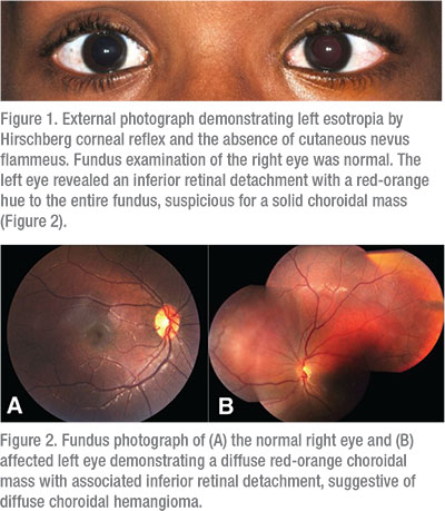
Ocular examination demonstrated visual acuity of 20/20 OD and 20/400 OS (pinhole to 20/200). Pupils were equal, round and reactive to light, with no relative afferent pupillary defect. Extraocular eye movements were full bilaterally. The patient demonstrated 20 prism diopters of left esotropia in primary gaze. Applanation tonometry revealed intraocular pressures of 10 mmHg in each eye. External examination was normal without birthmark or nevus flammeus (Figure 1). Slit lamp exam demonstrated few conjunctival papillae and mild constitutional melanosis, bilaterally.
Please click this link for diagnosis, workup, treatment and discussion.


