 |
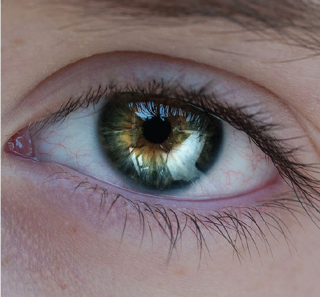
Researchers at the University of Hong Kong have found that SARS-CoV-2, the cause of COVID-19, replicates in the conjunctiva to a greater extent than the severe acute respiratory syndrome coronavirus that triggered the notorious SARS epidemic (SARS-CoV) that afflicted 26 countries with more than 8,000 cases in 2003.1 In fact, one of the researchers, in an interview with the South China Morning Post, says SARS-CoV-2 is much more efficient in infecting the human conjunctiva and the upper respiratory airways than SARS, with virus level about 80 to 100 times higher, which might explain its high transmissibility. However, the researchers and a leading corneal specialist in the United States, emphasize that the findings don’t necessarily translate to an increased risk of transmission of COVID-19 through the conjunctiva.
In a study published in Lancet Respiratory Medicine on May 7, the researchers isolated SARS-CoV-2 from a patient with confirmed COVID-19 and compared virus tropism and replication competence with SARS-CoV, Middle East respiratory syndrome-associated coronavirus (MERS-CoV), and 2009 pandemic influenza H1N1 (H1N1pdm, Swine Flu) in ex vivo cultures of human bronchus (n=5) and lung (n=4). The researchers also assessed extrapulmonary infection using ex vivo cultures of human conjunctiva (n=3) and in vitro cultures of human colorectal adenocarcinoma cell lines.
“This study shows what has been suspected—that conjunctival tissue can be infected with the SARS-CoV-2 virus,” says Yuri McKee, MD, MS, a corneal and refractive surgeon at East Valley Ophthalmology in Mesa, Arizona. “In general, I don’t think this is a significant source of transmission, especially as compared to airborne droplets from the respiratory system. Most personal protective equipment worn includes a mask and eye protection. This study didn’t show that that transmission via the eye was by the airborne route. Instead, it shows that the conjunctiva can be a source of infection. The most likely vector for conjunctival infection is probably fingers near the eyes.”
Kendrick C. Shih, MD, an ophthalmologist and one of the authors of the study, says his team’s ex vivo research model was based on the group’s previously published work comparing the differential ability of the 2009 H1N1 virus and the seasonal H1N1 virus to infect the conjunctiva.2 “We believe this finding represents a first step into better understanding how SARS-CoV-2 invades and replicates itself in the conjunctiva,” he adds. He recommended using “extreme caution” in translating the results into clinical practice, noting that other factors weren’t modeled in the ex vivo system, including the effect of the eyelids, blinking and the tear film. “We also don’t have evidence from our ex-vivo model that the virus spreads to the rest of the body through the blood stream,” he continues. “Finally, the study doesn’t examine whether the virus can then be spread from the conjunctiva of COVID-19 patients to others through tears, or through eye rubbing.”
Dr. Shih adds that the study further supports the idea that conjunctiva may be a potential tissue through which SARS-CoV-2 enters the body and that its replication is 40-fold higher than that of SARS-CoV. “But there is little evidence from our work and other studies to support the conjunctiva as a route of spread of the virus to others,” he notes. In fact, he adds, a recent clinical study conducted in Singapore demonstrated a significantly low SARS-CoV-2 RNA yield from the tears and conjunctival swabs of COVID-19 patients.3 “To answer this question, we will require further clinical and laboratory-based studies for better evidence of transmission through the conjunctiva,” says Dr. Shih.
Dr. Shih and his research colleagues note that ophthalmic care in Hong Kong is a “particularly high-risk area for spread of the virus” because of close face-to-face contact during exams, general overcrowding of public waiting areas and a potential risk of viral inoculation through the conjunctiva. “Ophthalmic centers have been amongst the first in stepping up precautions in the early days of the pandemic,” he continues, adding that plastic barrier shields have been installed to provide protection at slit lamps. “Our group recently shared with ophthalmologists across the world our experience in maintaining our routine ophthalmic clinic service, as well as our elective cataract surgery service.”4,5
Activities that are risks for generating micro-aerosols—including non-contact air-puff tonometry and those that involve tear, fluid or blood spillage, such as nasolacrimal duct syringing and incision and curettage of chalazia—were temporarily halted by clinicians in Hong Kong. Nursing staff assigned to apply eye drops for pupil dilation or topical anesthesia were at risk for splashes and were required to wear eye shields while performing these tasks, he adds. “With these measures in place, our clinics did not record a single case of COVID-19 infection amongst our staff and patients from the beginning of our introduction of precautionary measures on January 29 until April 19 of this year.”
1. Hui KP, Cheung MC, Perera RAPM, et al. Tropism, replication competence, and innate immune responses of the coronavirus SARS-CoV-2 in human respiratory tract and conjunctiva: An analysis in ex-vivo and in-vitro cultures. Lancet Respir Med 2020; MAY 7 . [Epub ahead of print].
2. Chan, M.C., Chan RWY, Yu WCL, et al. Tropism and innate host responses of the 2009 pandemic H1N1 influenza virus in ex vivo and in vitro cultures of human conjunctiva and respiratory tract. Am J Pathol. 2010;176:4:1828-40.
3. Seah IYJ, Anderson DE, Kang AEZ, et al. Assessing viral shedding and infectivity of tears in coronavirus disease 2019 (COVID-19) patients. Ophthalmology 2020; March 24. (Epub ahead of print].
4. Shih, C.K., Chan J.C.H. and Lai, JSM. Maintenance of ophthalmic specialist out-patient service during the COVID-19 outbreak: The University of Hong Kong experience. Eye (Lond) 2020; April 21. [Epub ahead of print].
5. Shih, KC, Wong JKW, Lai JSMet., Chan JCH. The case for continuing elective cataract surgery during the COVID-19 pandemic. J Cataract Refract Surg 2020; May 1. [Epub ahead of print].
AI Drusen Detection for Prediction of AMD Progression
Ophthalmologists at the Medical University of Vienna note that morphologic changes and their pathognomonic distribution in the progression of age-related macular degeneration aren’t well understood. In an attempt to get a better grasp on these changes, they undertook a study to characterize the pathognomonic distribution and time course of morphologic patterns in AMD, and to quantify changes associated with progression in macular neovascularization and macular atrophy.1
The cohort study included optical coherence tomography volumes from study participants with early or intermediate AMD in the fellow eye in the HARBOR (A Study of Ranibizumab Administered Monthly or on an As-needed Basis in Patients With Subfoveal Neovascular Age-Related Macular Degeneration) trial. Individuals underwent imaging monthly for two years (July 1, 2009, to Aug. 31, 2012) following a standardized protocol. Data analysis was performed between June 1, 2018, and Jan. 21, 2020.
To obtain topographic correspondence between individuals and over time, investigators mapped scans into a joint reference frame. They established time of progression to MNV and MA, and automatically segmented drusen volumes and hyperreflective foci (HRF) volumes in three dimensions using validated artificial intelligence algorithms. They constructed topographically-resolved population means of the markers by averaging quantified drusen and HRF maps in the patient subgroups. Here are some of the findings:
• Of 1,097 individuals enrolled in HARBOR, 518 had early or intermediate AMD in the fellow eye at baseline (mean age: 78.1 ±8.2 years; 59.7 percent female).
• During the 24-month follow-up period, 135 eyes (26 percent) developed MNV, 50 eyes (10 percent) developed MA and 333 eyes (64 percent) didn’t progress to advanced AMD.
• Drusen and HRF had distinct topographic patterns. Mean drusen thickness at the fovea was:
— 29.6 µm (CI, 20.2 to 39 µm) for eyes progressing to MNV;
— 17.2 µm (CI, 9.8 to 24.6 µm) for eyes progressing to MA; and
— 17.1 µm (CI, 12.5 to 21.7 µm) for eyes without disease progression.
• At 0.5-mm eccentricity, mean drusen thickness was:
— 25.8 µm (CI, 19.1 to 32.5 µm) for eyes progressing to MNV;
— 21.7 µm (CI, 14.6 to 28.8 µm) for eyes progressing to MA; and
— 14.4 µm (CI, 11.2 to 17.6 µm) for eyes without disease progression.
• The mean HRF thickness at the foveal center was 0.072 µm (CI, 0 to 0.152 µm) for eyes progressing to MNV, 0.059 µm (CI, 0 to 0.126 µm) for eyes progressing to MA and 0.044 µm (CI, 0.007 to 0.081) for eyes without disease progression.
• At 0.5-mm eccentricity, the largest mean HRF thickness was seen in eyes progressing to MA (0.227 µm; CI, 0.104 to 0.349 µm), followed by eyes progressing to MNV (0.161 µm; CI, 0.101 to 0.221 µm) and eyes without disease progression (0.085 µm; CI, 0.058 to 0.112 µm).
Investigators found that drusen and HRF represented imaging biomarkers of disease progression in AMD, demonstrating distinct topographic patterns over time that differed between eyes progressing to MNV, eyes progressing to MA and eyes without disease progression. They added that automated localization and precise quantification of these factors may help to develop reliable methods of predicting future disease progression. REVIEW
1. Waldstein SM, Vogl WD, Bogunovic H, et al. Characterization of drusen and hyperreflective foci as biomarkers for disease progression in age-related macular degeneration using artificial intelligence in optical coherence tomography. JAMA Ophthalmol 2020; May 7. [Epub ahead of print].
 |


