Just two or three years ago, if you were interested in using a femtosecond laser to make your LASIK flaps, you only really had to learn about one device, the IntraLase FS. Now, however, there are three additional femtosecond lasers approved in the
The AMO/IntraLase FS
The FS, often referred to as simply the IntraLase laser, has become the de facto standard by which any subsequent femtosecond laser is judged, simply because it was here first and many surgeons trust it to make consistent flaps. Here is a review of its operation.
In the beginning of the IntraLase flap-making procedure, after the surgeon applies anesthetic, he affixes a vacuum ring to the eye. "The main unique aspect of the ring is that it uses silicone skirting that requires less vacuum to attain applanation than the average microkeratome and is a little more comfortable for the patient," says Brian Will, MD, of 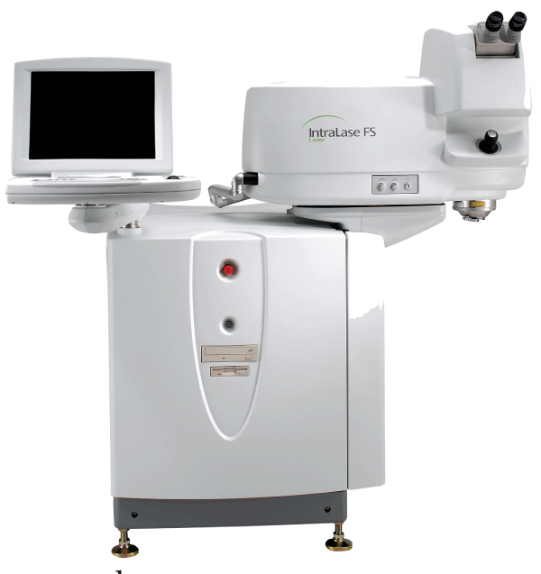
The next step is to bring the sterile, disposable docking cone, which mounts on the head of the laser, down onto the eye. This step has changed over the years as surgeons have become more familiar with the idiosyncrasies of making a femtosecond flap. "We originally did a wall-to-wall applanation on the cornea," says Dr. Will. "With this, you basically applanated it so the area applanated on the cornea was equal to the cone's surface area. However, I found that this excessively compressed the tissue, creating a situation in which you've already got the IOP raised and then you come along and flatten the cornea as well in a way reminiscent of using a vise. Now, however, the goal is 'soft docking,' which I described in 2002. In this approach, you use the smallest amount of applanation pressure possible to create the flap while optimizing the amount of applanation area so it's equal to or slightly larger than the area of the cornea you want to make the flap from. This allows you to leave an unapplanated meniscus around the edge. The advantage is you use less pressure and create less tension in the cornea, which is beneficial in minimizing the deleterious effect of the laser on the tissue, allowing room for the expanding fluid and gas created by the photodisruption to accumulate in the meniscus, rather than having it expand into the tissue and corneal lamellae."
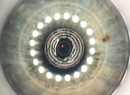
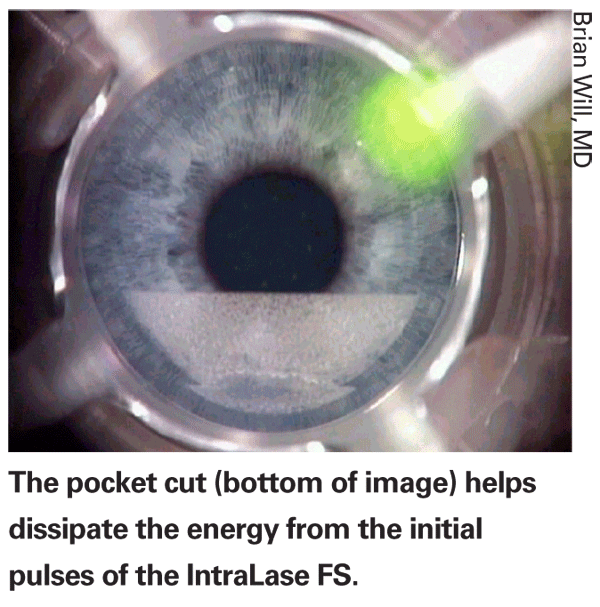
The IntraLase makes use of a corneal pocket cut, made at a depth of around 220 µm and with dimensions of 0.5 mm by 2 to 3 mm, to receive the expanding gas and fluid from its initial pulses. Along with the pocket, Dr. Will says surgeons have also markedly reduced the energies used by the laser to lessen the impact of these first pulses. The energy decrease was made possible due to the recently increased pulse rate of 60 kHz, up from 30 kHz. Using a flat applanation may also help blunt the effect of these pulses. "The flat applanator pushes the effects of the 'initial bang' out to the periphery of the cornea, to mask a lot of its adverse effects," says Dr. Will.
For energy delivery, the IntraLase uses a raster scanning pattern that scans in straight lines across the cornea, as opposed to a spiral pattern. However, for some of its corneal transplant applications, a spiral is possible, as well. The energy used for flap creation has decreased greatly over the IntraLase laser's life, from the 5 µJ used in 2002 to 0.7 to 0.8 µJ used today.
The IntraLase allows the surgeon to alter the following flap-creation parameters:
• flap diameter;
• side-cut angle and separation of the side-cut layers (i.e., the increments, in microns, that the device moves up as it cuts the side opening of the flap);
• spot/line separation of the raster pattern in microns (i.e., how far apart each spot is in a line/how far apart each line is from the next line; Dr. Will usually uses an 8-µm separation);
• energy per pulse;
• depth of pocket;
• number of pulses in the pocket;
• position and width of the hinge; and
• flap thickness.
In terms of flap thickness, Dr. Will says, at this stage of the technology, 100 µm is the thinnest flap the laser can cut very reproducibly and with a good level of safety. Though the estimates of standard deviation have varied, he says that, "after making these flaps day after day 'in the trenches,' " the flaps are usually within 10 µm of the target thickness. "I wouldn't recommend going thinner than 100 µm," he says. "To do that, you'd have to have an optical coherence tomographer to measure each flap as you cut it to see exactly what you're getting each time."
Ziemer's Femto LDV
The Femto LDV already has about 20 lasers in place in the
Though the Femto LDV uses flat applanation like the IntraLase, instead of having a laser cone that docks with the eye, the Femto LDV puts the laser optics in a handpiece that sits on the end of an articulated arm that can be swung into and out of the operative area. "You control the applanation manually, like a microkeratome," explains Anton Wirthlin, PhD, Ziemer's vice president of marketing. The arm can be moved out of the way to allow the excimer microscope to be brought in, rather than having to move the patient to do the excimer step. Dr. Wirthlin says the company achieves applanation with less suction than a microkeratome, though experiments to determine just how much pressure is exerted on the eye are ongoing. The system uses computer-controlled suction in a way similar to the Amadeus microkeratome. "The computer regulates the suction," explains Philips Kirk Labor, MD, of Grapevine,
One aspect of the Femto cut that takes some getting used to is the fact that it's a blind cut. The surgeon's view is completely obscured by the handpiece's optics as the flap is being made. "Yes, I had to adjust to that," says Dr. Labor. "But, when I used to use the Hansatome, I couldn't see the cut then, either. You do have to get used to it, but it's not something I'm completely unaccustomed to." The system does provide the option of interrupting the cut to take a peek at how things are progressing, however.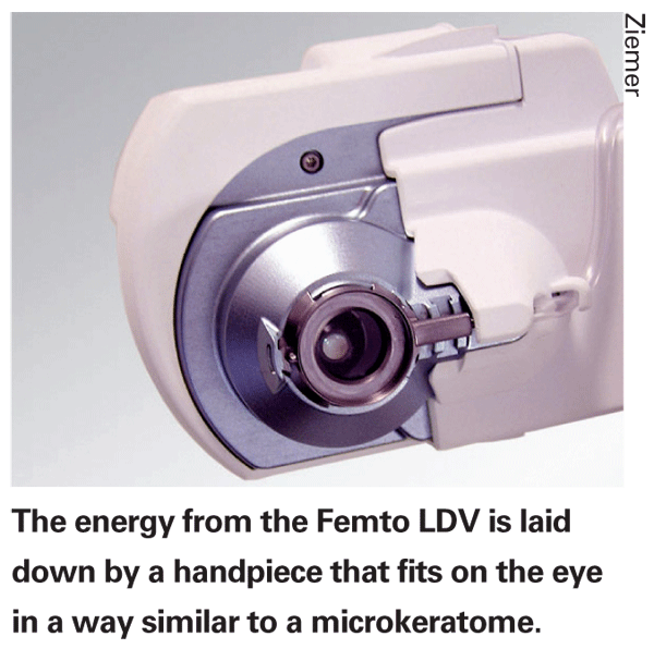
The flap thickness is determined by a sterile device placed in the applanation window of the laser called an Intershield. To cut a certain depth, you need a certain Intershield. "When we first used the device, as can happen the first time with anything, we had a couple of patients with a couple of areas on the flap that were hard to separate from the bed," recalls Dr. Labor. "We think it had to do with the Intershield. If it's not completely clean and there's a little spot on it, the laser light can't traverse that spot and you'll get a spot that's uncut. So now, we make sure the Intershield is very clean to begin with." Flap diameter is determined by the size of the suction ring that's used, and rings range from 8.5 mm to 10 mm in 0.5-mm increments. The hinge location can't be placed just anywhere the surgeon would like. Instead, it needs to be placed at one of the four cardinal directions. However, the surgeon can alter that by about 30 degrees clockwise or counterclockwise by turning the handpiece.
The Femto LDV uses an extremely fast pulse rate, around several thousand kHz (Ziemer declines to reveal the actual speed). "This isn't meant to imply that the cutting process is faster than, say, the IntraLase," says Dr. Wirthlin. "Instead, we focus the beam very tightly so that we can get away with very minimal pulse energy, a few nanojoules per pulse rather than microjoules. The result is we have to place many more spots that are closely spaced. The spot size is a few microns wide and the spots are tightly overlapped, resulting in a smoothly cut surface without tissue bridges between spots [that would need to be broken to lift the flap]." Flap creation can take between 20 and 35 seconds.
So far, Dr. Labor says he hasn't experienced any corneal inflammation with the device. "The cuts have been clean and I haven't had diffuse lamellar keratitis," he says.
The Femto LDV is also portable, both in the sense of putting it in a truck and taking it somewhere to do surgery, and in the sense of just moving it around your office. "Since the normal position for the Femto LDV is on the left side of the bed, with the excimer on the right, in order to make it easier for patients to lie down and get off the bed, some surgeons will pull the Femto LDV away from the bed and then push it back into place again using its wheels," says Dr. Wirthlin. "You can do this without any danger of damaging it or putting it out of alignment."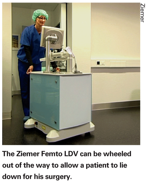
Dr. Wirthlin says the company's experience in the
Carl Zeiss Meditec's VisuMax
The VisuMax is the femtosecond laser most recently approved by the U.S. Food and Drug Administration. It features a curved glass applanator to preserve the corneal shape, low-pressure docking and the ability to perform an all-femtosecond refractive procedure (currently awaiting CE Mark approval in
"The VisuMax, like the [20/10 Perfect Vision] Femtec, has a curved contact glass that doesn't change the corneal curvature that much," says Walter Sekundo, MD, medical monitor of the VisuMax's European clinical studies. "This is a huge advantage if you want to do refractive treatments with your femtosecond laser, as is now possible with the VisuMax."
The VisuMax cuts at a 200-kHz frequency, uses an energy density just below 300 nJ and uses a spiral cut pattern as opposed to a raster. Dr. Sekundo says the spot separation is between 3 and 6 µm, depending on the laser mode in which you're operating.
"In the laser's 'Fast' mode, the spot separation is a little larger so the entire cut is much faster than if they were closer together," explains Dr. Sekundo. "In Fast mode, I need around 21 seconds to make the flap. Though the frequency is faster than the IntraLase, both lasers take about the same time to make a flap because the VisuMax has to make a cut on a curved surface rather than a flat one." He says there's also an "Enhanced" mode that gives the option of increasing the energy used in cases where you want to make the flap easier to lift.
Carl Zeiss Meditec is also touting the VisuMax's ability to achieve suction without causing the patient's vision to black out. "It's a different concept of suction on the globe compared to other lasers. The applanation is on the surgical limbus and cornea, and there's no conjunctiva involved," explains Dr. Sekundo. "If you're on conjunctiva, you have to suck in the conjunctiva first and need much higher suction to then achieve suction on the sclera. The VisuMax puts a cup-like device on the eye rather than a glass plate that squeezes the cornea in a sandwich-like manner. You basically move the patient toward this cup. And once the applanation is there, you push the pedal, the suction starts and the laser is ready."
The selectable flap parameters include flap diameter in 0.1-mm increments, the location of the hinge, the flap thickness and the side-cut angulation. Dr. Sekundo thinks that the thinnest flap that surgeons should probably routinely cut with the laser is between 100 µm and 110 µm thick, depending on the cornea.
A unique attribute of the VisuMax is its ability to perform an all-femtosecond refractive procedure. The procedure, called Femtosecond Lenticule Extraction (FLEX) involves just what its name implies: The VisuMax intrastromally dissects a lenticular piece of corneal tissue, then creates a flap as a single-step procedure. The refractive lenticule is then manually removed by the surgeon. This extraction creates the refractive effect. Dr. Sekundo's colleague, Marcus Blum, MD, presented the interim results of their FLEX study of 45 patients at the 2007 meeting of the European Society of Cataract and Refractive Surgeons. The mean preop spherical equivalent refraction was -4.43 D, with less than 6 D of astigmatism. The surgeons removed lenticules between 6 and 7.3 mm in size, depending on the patient's mesopic pupil size, and always left more than 300 µm of stromal bed postop. The target refraction was between
At six months postop, the average refractive error was +0.25 D, and 66 percent of the patients were within ± 0.5 D of the intended postop refraction. Seventy-one percent had a visual acuity of at least 1.0 (20/20 on the Snellen chart).
In terms of adverse events, the surgeons reported one case of mild haze, several cases of microstriae and four cases in which suction was lost and the patients were retreated with LASIK or LASEK. "FLEX will receive a CE Mark soon, and we'll then start these as normal treatments, rather than just investigative ones," says Dr. Sekundo.
20/10 Perfect Vision's Femtec
The Femtec is the other femtosecond laser approved in the 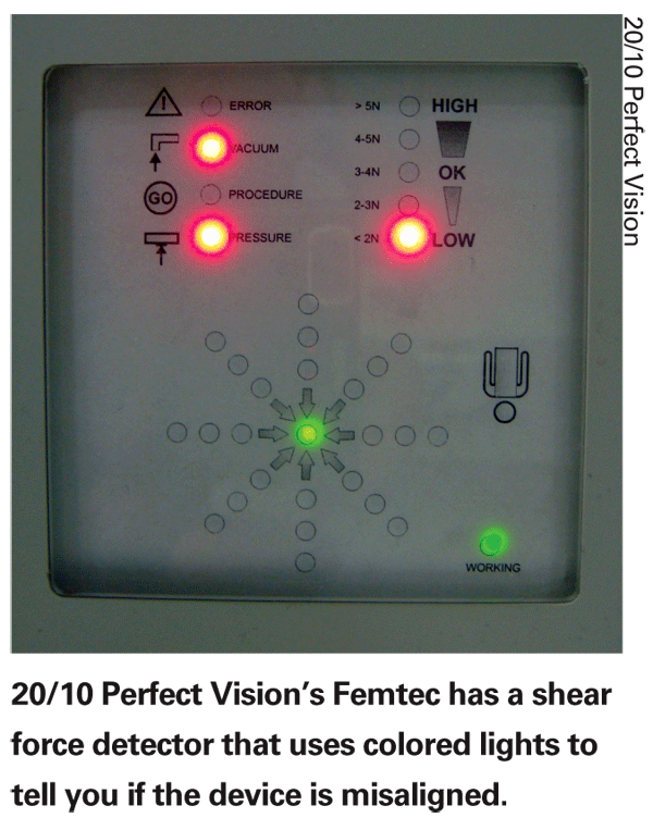
Mr. Behrend says the procedure does increase the pressure in the eye, but not as much as a flat applanation would. "We stay in the 20- to 35- mmHg range in IOP," he says. "That's a comfort to the patient because he won't experience the traditional blackout associated with microkeratomes, and won't have the other stresses put on the central retinal artery due to increased IOP." Despite the lower amount of suction, he says the device's users don't experience any more suction breaks than anyone else. "It's a rare phenomenon," he says.
Another feature aimed at safer docking is the device's radial shear detector. With this, sensors on the docking cone measure the precise placement of the eye and display the quality of the connection in a way similar to a traffic light. "You've got a green light for 'Perfect, go,' says Mr. Behrend. "Then you've got yellow for, 'It's OK, you can still go,' a slightly deeper orange for 'OK,' and a red light that communicates, 'You're off and have to re-adjust.' "
The Femtec laser engine is currently running at 40 kHz, and the surgeon can use anything from 850 to 1,500 nJ for making a flap, depending on the optics in the particular laser, says Mr. Behrend. For making the side cut, which Femtec calls the rim cut, it uses anywhere from 800 nJ for a LASIK flap cut to as high as 2,000 to 3,000 nJ for penetrating keratoplasty, which goes much deeper. "The physician can change rim angle; flap thickness; flap diameter (from 6 to 9.5 mm) and spot spacing," adds Mr. Behrend. "There's actually no option that can't be changed if the surgeon wants to try something different."
Mr. Behrend says that the company recommends going for between 100- and 130-µm flaps. The standard deviation in flap thicknesses is 10 µm. The spot spacing is surgeon selectable, and the usual spacing is between 5 and 12 µm.
To help decrease the energy that's put down, the Femtec has a double-pass pattern as an alternative to the usual single pass. "With this, the laser pattern goes twice with a much wider spacing," says Mr. Behrend. "This is advantageous because the bubbles that are created with the pulses will be more broadly spread apart, so there will be less energy going on the eye per square millimeter."
Though the Femtec isn't for sale in the