 |
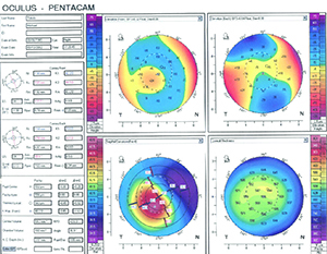 |
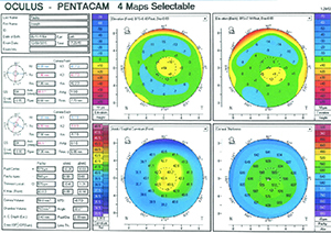
| When an eye has undergone previous hyperopic LASIK, an intraocular lens with positive spherical aberration may help to improve the patient’s vision—if the ablation is well-centered (above, top). However, hyperopic ablations are often decentered; then the positive asphericity could backfire and a zero-aberration lens would be preferable (bottom scans). (Image courtesy Eric D. Donnenfeld, MD.) |
Spherical Aberration
As every cataract surgeon knows, determining the ideal power for an intraocular lens can be challenging in eyes that have previously undergone refractive surgery. But today, that’s not the only concern; modern lens technology has made it possible to address the spherical aberration of the cornea, which may have been altered by that surgery. Many surgeons try to counteract the alteration by choosing an IOL with a level of positive or negative asphericity that may offset it. (In fact, this strategy is now common-ly used to try to produce the best possible vision in eyes with virgin corneas, which normally have some spherical aberration as well.)
“Intraocular lens design has been one of the great innovations in ophthalmology over the past decade,” says Eric D. Donnenfeld, MD, clinical professor of ophthalmology at New York University Medical Center and a partner at Ophthalmic Consultants of Long Island. “Previous generations of lenses had positive spherical aberration that added to the net spherical aberration of the cornea, resulting in patients having significant higher-order aberrations. Those aberrations led to glare and halo, a loss of contrast sensitivity and an overall loss of vision quality.
“The advent of new-generation lenses occurred about 10 years ago when Pharmacia developed the aspheric IOL, which has negative spherical aberration,” he continues. “The negative spherical aberration offsets the positive spherical aberration of the average cornea, resulting in better contrast sensitivity. These lenses achieved New Technology IOL status from the Centers for Medicare & Medicaid Services and were reimbursed at a higher rate than other lenses because it was proven that they improved people’s ability to function in tests such as driving ability.
“Today, these lenses are commonplace, and there are several different lenses that are available,” he says. “There are negative spherical aberration lenses, low-negative spherical aberration lenses, zero spherical aberration lenses and positive spherical aberration lenses. All of these play a role in my surgical armamentarium when managing patients undergoing cataract surgery. For routine cases, I generally choose a negative spherical aberration lens, unless the eye is unusual; for the overwhelming number of patients, negative spherical aberration lenses yield better quality of vision. But for special circumstances, having the full armamentarium of lenses available is helpful for matching the patient’s preoperative status and achieving the best postoperative surgical result.”
Managing Altered Sphericity
Dr. Donnenfeld notes that hyperopic LASIK leaves the cornea steeper, inducing negative spherical aberration. “For these patients I would either implant a zero-aberration lens or a positive spherical aberration lens,” he says. “If the topography shows that the center of the cornea is steepest along the line of sight, a positive spherical aberration lens can actually give better quality of vision. However, if it’s decentered, which commonly occurs with hyperopia, a zero aberration lens makes the most sense. (See examples, right.) On the other hand, when patients have had previous myopic LASIK their corneas are flattened, inducing positive spherical aberration. As a result, they generally have more positive aberration than a normal cornea. In this situation it’s very important not only to use a negative spherical aberration lens, but to use a lens which has the maximum amount of negative spherical aberration.”
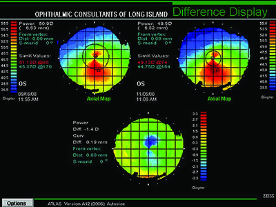 |
| Keratoconic eyes generally have negative spherical aberration and decentered optical apexes, making a zero-spherical aberration lens most appropriate. If the disease is mild-to-moderate (as in the case above) and the eye has some astigmatism, but not a significant amount of irregular astigmatism, a toric lens may improve vision. (Image courtesy Eric D. Donnenfeld, MD.) |
“If the patient was treated for hyperopia, the eye will now have negative spherical aberration,” he continues. “In these cases, if the correction was well-centered, I prefer to use a lens with positive spherical aberration—i.e., a conventional spherical lens. On the other hand, if the eye has undergone myopic LASIK, with good centration and no significant amount of coma, then I prefer to use negative asphericity lenses to compensate for the positive spherical aberration these patients usually have.” (Dr. Alio notes that eyes that have undergone refractive surgery within the past two or three years may not have this altered corneal spherical aberration because of improvements in the software used to perform the surgery. However, the majority of patients currently undergoing cataract surgery will have had any refractive surgery performed more than three years ago.)
“Another concern when patients have had previous corneal refractive surgery is toricity,” Dr. Alio says. “If the corneal topography shows that the patient has more than 2 D of corneal astigmatism, my choice is a toric lens, with either negative or positive asphericity, depending on the centration of the previous corneal refractive surgery. Toric lenses are an excellent choice for these patients because you address both the astigmatism and the spherical component.”
However, some surgeons’ experience has made them skeptical about the significance of the sphericity of the lens. “I think it’s uncertain how much benefit patients actually get by choosing a certain degree of asphericity in the IOL,” says Richard Mackool, MD, director of the Mackool Eye Institute and Laser Center and senior attending surgeon at The New York Eye and Ear Infirmary. “About 15 years ago when aspheric IOLs first became available, I was doing about 3,500 cataract surgeries a year and I had a lot of patients who had only had one eye done, using a standard IOL. Because the new technology was now available, I put an aspheric IOL in the second eye of about 500 patients. Not one of those patients noticed a difference. Even in the exam lane when we asked which eye was better, they couldn’t tell.
“Of course this was in normal, ambient lighting conditions where the pupil was relatively small,” he says. “But none of them noticed a difference driving at night, either. I’m aware that people have conducted tests that found that reflex stopping distance improved with an aspheric lens when driving in the dark, but no patients came in and told me that.
“It’s good that we keep searching for better IOLs, including higher-order-aberration-correcting IOLs,” he adds. “But thus far, my experience has been that to minimize problems caused by higher-order aberrations, it’s far more important for the patient to have a small pupil.” (For more on that topic, see the sidebar on p. 30.)
Keratoconus
One of the issues presented by keratoconic eyes is that they are exceptionally steep, causing negative spherical aberration. “Adding additional negative spherical aberration would not be a good idea,” Dr. Donnenfeld points out. “I prefer to use a zero spherical aberration lens in these patients because these eyes also have decentered optical apexes. A zero-aberration lens ensures that if the lens decenters from the line of sight it will not induce higher-order aberrations such as coma.”
In terms of implanting a toric lens, he notes that it’s impossible to fully correct an irregular cornea with a toric lens. “Toric lenses are optimal for patients who have regular bow-tie astigmatism,” he says. “However, in general, toric lenses do well when the patient has mild-to-moderate keratoconus. (See example, above.) I generally place the lens on the keratometric axis, but I like to confirm it with the refraction as well. On the other hand, if the patient has significant irregular astigmatism, a toric lens is generally a bad idea.”
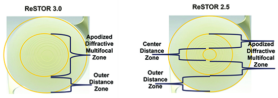 |
| Although most surgeons are hesitant to implant a multifocal lens in an aberrated eye, some recent multifocal lenses have reduced the potential for problematic outcomes by making the center area of the lens distant-dominant, as in the ReSTOR 2.5 (pictured above, right). Where previous multifocals could cause a 20-percent reduction in distance acuity, surgeons report that this format may only reduce the patient’s distance acuity by 5 percent. (Image courtesy Richard Mackool, MD, PC.) |
Dr. Mackool points out that typical cataract patients with keratoconus have a stable cornea because of their age; that frees the surgeon from having to worry about worsening keratoconus after implantation. “Most of these patients have significant astigmatism, and many have a lot of irregular astigmatism,” he says. “So, you’re probably not going to want to implant a multifocal—although a few of the newest multifocals might work.”
Dr. Mackool also offers some advice about determining the best refractive power for the implant. “Determining the IOL power can be very difficult because of the irregularity and multiple curvatures in the cornea,” he notes. “Conducting an aphakic refraction helps, but part of the problem in these cases is that the dilated refraction and normal-sized pupil refraction can be very different; when the pupil is dilated the light is entering the eye through far more irregular curvature. For that reason, the aphakic refraction really should be done not while the patient is dilated immediately postop, but the next morning when the patient has a normal pupil.
“For that reason, when we have a patient with any significant keratoconus we divide the operation into two parts,” he says. “On the first day we take the cataract out and perform an aphakic refraction; the next day we put in the implant. In fact, we do this with every patient in whom we can’t measure the IOL strength accurately by the usual means, including post-LASIK eyes and eyes with nystagmus. If we find significant cylinder, we’ll often use a toric IOL, but we will seldom use a multifocal IOL in these patients. We won’t use a toric IOL if the plan is for the patient to wear a hard contact lens after surgery.”
Dr. Mackool notes that some surgeons express concern about dividing the surgery into two parts. “People say we’re doubling the risk of infection by doing this, but we’ve had zero infections and zero complications in more than 2,000 patients,” he says. “Of course, part of the reason is that we put vancomycin in the eye at the end of the procedure. We’ve done 80,000 consecutive cataract surgery operations at my surgery center without one case of endophthalmitis. There was a recent report of possible retinal vasculitis after intracameral vancomycin injection in the literature, but we’ve used vancomycin in 80,000 consecutive cases at our center with no en-
dophthalmitis and no sign of any type of postoperative vasculopathy.”
Multifocal Contraindications
Many surgeons avoid implanting a multifocal IOL whenever an eye has any unusual condition. However, according to Dr. Mackool, the design of some recent multifocal IOLs has made them less problematic in non-standard eyes. “Some new IOLs under development, including the ReSTOR 2.5, which is already available, are distant-dominant in the central 1 mm,” he says. “This alleviates much of the concern we’ve had about implanting a multifocal in an aberrated eye, whether the problem is keratoconus or some other disease that causes corneal irregularity. I’ve used it successfully in these patients many times.”
Dr. Mackool says the reason this can work in an aberrated eye is the way the light is distributed by the lens. “It comes down to the percentage of light rays devoted to distance, especially in the center of the lens,” he explains. “If an eye already has some issues with focusing 100 percent of light rays appropriately—e.g., keratoconus or post-LASIK—now you may be talking about a multifocal that’s using half of the light rays for distance and half for near. This can lead to one very unhappy patient. However, if the IOL devotes the vast majority of light rays to distance because the central millimeter is 100-percent dedicated to distance, this is far less problematic.
“Prior to the availability of the ReSTOR 2.5 lens, I would tell my patients who were going to receive a multifocal lens that their distance vision would be about 20-percent reduced,” he says. “That would dissuade a lot of people from choosing a multifocal. But with the ReSTOR 2.5, the patient may lose only 5 percent of his distance acuity. That’s insignificant to virtually everybody. I wouldn’t implant it in a professional baseball player, but that’s about the only exception.”
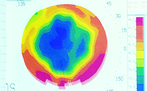 |
| A cornea that previously underwent radial keratotomy (example above) is likely to be left with multiple aberrations. Surgeons often choose a neutral-asphericity lens for these eyes, in part to avoid combining asphericity with aberrations such as coma, which can be problematic. A neutral lens is also less problematic if the ablation or lens is decentered. (Image courtesy Eric D. Donnenfeld, MD.) |
“I tend to be conservative about placing multifocal lenses in patients who have irregular corneas,” adds Dr. Donnenfeld. “The most common irregular cornea we see is one that has undergone previous laser vision correction. If the cornea looks normal and topography shows a well-centered ablation with a modern ablation zone that’s fairly prolate, then a multifocal lens may work pretty well. But if the cornea has an older-style ablation profile, especially in patients who started out with high myopia or hyperopia, I avoid implanting multifocal lenses because of the likelihood of a loss of contrast sensitivity.”
Weak Zonules
“If a patient has weak zonules or there’s concern about IOL decentration, a zero-aberration lens is my lens of choice because of the possibility of the IOL moving off of the visual axis,” notes Dr. Donnenfeld. “A patient who has had a previous vitrectomy, for example, will often have weak zonules. You just have to prepare for that when you operate on the patient.
“Surgeons have different opinions about the best lens to implant when zonules are weak,” he continues. “Some surgeons like to use one-piece acrylic lenses because placing them in the eye is less traumatic. Some people like three-piece lenses because they provide a pseudo capsular tension ring, holding the capsular bag in place. In addition, if the lens does dislocate, it’s much easier to suture a three-piece lens to the iris than a one-piece lens. In general, I choose to use three-piece lenses in eyes that have poor zonular support, such as patients with pseudoexfoliation, patients who have undergone trauma, and occasionally patients who have had a vitrectomy.”
Dr. Alio agrees. “If the patient has had a vitrectomy, I usually implant a three-piece lens, the Alcon MA60, and I usually use a capsular tension ring,” he says. “In many cases these capsules are quite unstable, and in my hands this approach works better.”
Dr. Mackool notes that some surgeons believe toric or multifocal IOLs should not be used in eyes with poor zonular support, but he disagrees. “I’ve placed those IOLs in many of these eyes, typically pseudoexfoliation eyes with a lax zonule,” he explains. “People say, ‘What if the lens dislocates?’ Well, then you have to fix it, just as you’d have to fix an aspheric monofocal. The fact that you might have to reposition the lens at some point doesn’t decrease the patient’s chances of seeing well with a toric lens or premium IOL.
| The Pupil-Size Factor |
| Today, when a patient has had previous laser refractive surgery, many surgeons attempt to correct for spherical aberration that may be present in the cornea as a result of the surgery by choosing an implant with a specific amount of negative or positive spherical aberration. However, some surgeons believe this may not be necessary in many patients. “In reality, the single most important factor in terms of how well these patients do, especially in dim illumination, is their pupil size,” says Richard Mackool, MD, director of the Mackool Eye Institute and Laser Center and senior attending surgeon at The New York Eye and Ear Infirmary. “If the patient has a small pupil—the ideal pupil size is between 2 and 2.5 mm, as measured by the observer—then you pretty much don’t have to worry about the higher-order aberrations that may have been introduced by the corneal procedure. So it’s very important to measure the pupil size in these patients preoperatively. If the patient has a large pupil, then you should have a discussion about the possibility of using a miotic agent. “For example, I’ve seen several patients over the years with large pupils post-LASIK in whom we implanted IOLs, and their best-corrected visual acuity would be something like 20/50,” he says. “Then, we’d put in a miotic agent and take the pupil down to 2.5 mm, and they’d be 20/20. A number of those patients were referred to me with the idea that they might need an IOL exchange. Instead, I put them on miotics and they looked at me like, ‘Why didn’t somebody else think of this?’ I believe the answer is simply that this approach hasn’t been well-publicized.” He adds that a pupil size of 2 to 2.5 mm is optimal because it provides the best depth of field, while avoiding the problem of noticeably reduced vision in dim lighting conditions. Dr. Mackool notes that using a miotic has had some downsides in the past, but that may be about to change. “Until now, the only miotic agent we’ve been able to use has been pilocarpine,” he says. “A weak solution of about 0.5% is all one needs, but that means you must either obtain it from a compounding pharmacy or have the patient dilute it, because the lowest commercially available concentration is 1%. To minimize the expense and effort of obtaining it already diluted, we have patients use the 1% drops and put in a lubricating drop simultaneously, which roughly dilutes it down to 0.5%. Of course a few patients—about 10 or 15 percent of them—will need to use 1% pilocarpine because 0.5% doesn’t cause enough reduction in pupil size. “Unfortunately,” he continues, “using pilocarpine for this purpose can cause other side effects. It stimulates accommodation, although that’s not a problem after cataract surgery. It also gives some users a periorbital brow ache, especially the first few days they use it; that ache may continue for quite a while in some younger patients. A very small percentage of users will experience irritation or even develop a little iritis. Also, the maximum benefit you get out of pilocarpine only lasts about six hours, so the patient has to use it two or three times a day. However, this may change soon, thanks to a new miotic that promises to be much easier to use, one that reduces pupil size but doesn’t stimulate accommodation. Its activity is limited to the iris only. It promises to be much better tolerated, and will have a much longer duration of action. So, help is on the way. I can’t wait to get my hands on the new drop myself, because I’m tired of using reading glasses.” —CK |
Other Issues
Other factors may also make it advisable to consider implanting an IOL other than your standard choice:
• Patient age. Dr. Alio says the age of the patient does matter. “In cases of elderly patients, I don’t worry as much about asphericity,” he says. “These patients usually have small pupils and don’t benefit much from choosing a special spherical or aspherical lens. Also, standard spherical lenses are much less expensive, which can be important to these patients. It’s something to consider in patients over the age of 75.
“I also avoid using multifocal lenses in patients over 75,” he adds. “The learning capacity of an aging brain is not as great as that of a young brain. Neural adaptation and contrast sensitivity both drop with age, and this becomes more significant when we reach our 70s. This is just the normal evolution of the human brain and eye and retina.”
• Patients wearing gas-permeable contact lenses. “Implanting a toric lens in a keratoconus patient who wants to continue to wear gas permeable contact lenses after cataract surgery is not a good idea,” says Dr. Donnenfeld. “Once the lens is placed you lose the effectiveness of the gas-permeable contact lens because the cylinder is placed inside the eye and can no longer be corrected with a contact lens.”
• Lens in the sulcus. Dr. Donnenfeld notes that poor zonular support is a common reason for placing the lens in the sulcus. “Under these circumstances, three-piece lenses are ideal,” he says. “In addition, the lens that’s placed in the sulcus should have a longer length. No IOLs in the United States are specifically designed for sulcus placement, but the STAAR AQ2010 and AQ 5010 lenses are the longest lenses currently available—13.5 and 14 mm, respectively. These are very helpful when the lens is in the sulcus, especially in larger eyes, where the white-to-white may cause helicoptering of a shorter lens.”
Dr. Donnenfeld adds that in most cases, it’s not a good idea to place a one-piece acrylic lens in the sulcus. “This will cause iris chafing and can induce uveitis-glaucoma-hyphema syndrome,” he notes. “However, an acrylic lens placed in the bag with optic capture anteriorly is a successful procedure, and it’s something that can be done for patients receiving a premium lens, such as a toric or multifocal lens, when there’s a rent in the posterior capsule.”
• Silicone vs. acrylic. Although silicone lenses work well in most situations, acrylic lenses are more popular today. “Typically, acrylic IOLs can be inserted through a smaller incision and they unfold more gently,” notes Dr. Mackool. “I think the vast majority of retinal surgeons favor an acrylic IOL in eyes that are at risk of retinal detachment, because if the patient may have retinal problems or retinal detachment you may need to use silicone oil inside the eye. You tend to get beading when you have silicone oil in contact with a silicone lens, so you might end up having to exchange the IOL. However, this is not common today.”
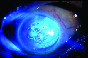 |
| An eye with Fuchs’ dystrophy. In this situation, some surgeons recommend implanting a lens that will leave the patient mildly myopic, because a possible future DSEK could induce a diopter of hyperopia. (Image courtesy Eric D. Donnenfeld, MD.) |
“In general, I prefer to implant acrylic hydrophobic lenses in eyes that have had a previous vitrectomy,” says Dr. Alio. “I never use silicone lenses. In my experience, silicone lenses are much more fragile, and they decenter more easily than the acrylic lenses. They don’t fit as well in the posterior capsule. And in cases of previous vitrectomy, many cases have had silicone oil in the eye; silicone with silicone doesn’t produce good results in the postoperative period.”
• Piggyback lens. “There are times when a patient has had previous refractive surgery and the best refractive outcome is achieved by adding a piggyback lens,” says Dr. Donnenfeld. “Under these circumstances it’s always best to have one lens in the bag and one lens in the sulcus. The lens in the sulcus, of course, should be a three-piece lens. In addition, it’s been found that it’s best if the two lenses are made of different materials. So if a silicone lens is in the bag, place an acrylic lens in the sulcus, and vice versa. When the piggyback lens is made of the same material there are interface issues; the result is always more inflammation.”
• Patients who have Fuchs’ dystrophy. “In these patients I like placing a lens that leaves the patient mildly myopic,” says Dr. Donnenfeld. “They may need a DSEK in the future, which could induce a diopter of hyperopia.”
• A family history of macular degeneration. “Many surgeons avoid using a multifocal in a patient with a family history of macular degeneration,” notes Dr. Mackool. “They’re thinking, ‘What if the patient gets macular degeneration down the road?’
“I don’t buy that reasoning,” he says. “It’s OK to use a multifocal in those patients, because first of all, you don’t know whether they’ll ever get macular degeneration. Second, medicine is likely to progress in the years ahead. Who knows what kind of therapies will appear in the next few years, therapies that may prevent them from getting the disease or at least keep it from becoming serious? And even if you do need to change the IOL at some point in the future, there may be new techniques for accomplishing that. Third, with a lens like the ReSTOR 2.5, which has far less impact on distance vision, the lens is much less likely to be an issue even if the disease does appear. So I don’t think it makes sense to limit your options too much because the patient might have a problem at some point in the future.” REVIEW
Dr. Mackool is a consultant to Alcon. Dr. Donnenfeld is a consultant for Alcon, Bausch + Lomb, Zeiss and Abbott. Dr. Alio has no financial ties to any product discussed.


