A 59-year-old male was referred to Wills Eye Hospital for evaluation of three months of bilateral blurred vision. Recent medical history revealed fatigue and weight loss necessitating inpatient admission, with an evaluation revealing a diagnosis of acute myeloblastic leukemia. At that time, his white blood cell count was 46,000 cells/mcL (normal: 4,500 to 10,000 cells/mcL); it peaked at 92,000 cells/mcL during his hospitalization. His course was complicated by admission to the intensive care unit for findings of leukostasis, acute respiratory distress syndrome and distributive shock that resolved with emergent leukophoresis. He was managed with induction chemotherapy using cytarabine and daunorubicin. He did not receive radiotherapy or surgery.
Medical History
Past medical history was significant for a 10-year history of type II diabetes mellitus, hypertension and hyperlipidemia. Prior to hospitalization, a local optometrist had diagnosed the patient with non-proliferative diabetic retinopathy.
Social history was unremarkable, but family history was significant for diabetes mellitus, renal cell carcinoma (sister) and colon carcinoma (brother). The patient’s medications included metoprolol, metformin and insulin. He had no known drug allergies.
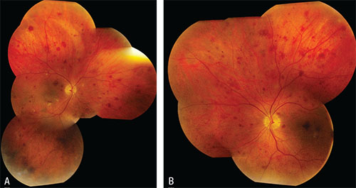 |
| Figure 1. Montage fundus photography of the right (A) and left (B) eyes demonstrating scattered dot and blot retinal hemorrhages in both eyes and circumpapillary and macular nerve fiber layer infarctions, predominantly in the right eye. |
Examination
On examination, uncorrected visual acuity was 20/150 with pinhole improvement to 20/50 in the right eye and 20/70 in the left eye. External examination was unrevealing. Intraocular pressure was 11 mmHg OD and 13 mmHg OS. The anterior segment was normal in each eye except for mild nuclear sclerotic lens opacity in both eyes.
Funduscopically, both eyes showed similar features of scattered dot and blot intraretinal hemorrhages involving the entire fundus (See Figures 1A, 1B). There was no evidence of leukemic infiltration in the retina, choroid, vitreous or optic disc. Occasional white-centered hemorrhages were noted in the macular region. In the circumpapillary region, there were several nerve fiber layer infarctions. A coincidental flat choroidal nevus was noted along the inferotemporal vascular arcade, measuring only 0.5 mm in diameter.
Please click this link for diagnosis, workup, treatment and discussion.


