Modern refractive surgery has been proven to be safe and effective time and again, but that doesn't stop researchers from attempting to hone its edge even further. This year's crop of refractive surgery abstracts from ARVO cover topics ranging from the corneal effects of various flap-making modalities to the shortcomings of epi-LASIK. Here are the latest results of this cutting-edge research.
LASIK Update
Researchers from
In this prospective study, the physicians performed bilateral LASIK on 40 patients with an average spherical equivalent preop refraction of -8.71 ±1.5 D (r: -6.31 to -12 D). Before surgery, they measured patients' pupils with a semi-automated, infrared pupillometer. At one year, patients completed a questionnaire on subjective visual quality before and after their surgery.
Postop, the mean spherical equivalent was -1.81 ±0.92 D (r: -0.06 to -3.81 D). LASIK caused no significant changes in best-corrected vision. Postop binocular uncorrected vision averaged 0.24 ±0.27 (between 20/32 and 20/40), with a range of -0.1 (20/16) to 1.0 (20/200). A year later, 72 percent of the patients say they agree or partly agree that they are very satisfied with their surgery. High satisfaction was correlated with a good postop uncorrected acuity (p=0.014), while self-reported quality of day or night vision had no significant impact on patient satisfaction. LASIK caused no significant changes in self-reported quality of vision in photopic conditions, but 64 percent of patients reported their night vision to be worse postop (p<0.001). A significant number reported an increase in glare, halos, double contours, "shadow" pictures and difficulty in seeing small details in scotopic conditions. Increased susceptibility to blinding by car headlights was reported by two-thirds of them (p<0.001). A large scotopic pupil correlated with reported glare (p=0.015) and blinding (p=0.026). Age, gender, photoablation depth or photoablation diameter didn't correlate with any of the reported visual disturbances.2901
Researchers from 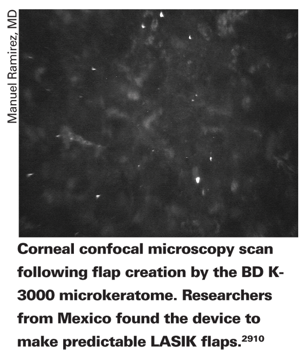
The physicians randomized 90 eyes of 45 patients to having a flap cut with the Bausch & Lomb Hansatome (to create a planned 160-µm flap), the BD K-3000 keratome (to create a 130-µm flap) or the IntraLase (to create a planned 100-µm flap). They scanned the corneal thickness with the Confoscan 4 confocal microscope (Fortune Technologies,
Only in the BD K-3000 group was the created flap thickness not statistically significantly different from the planned thickness (111.7 ±12.9 µm; p=0.137). The measured thickness in the Hansatome group was thinner than planned (146.5 ±20.3 µm; p<0.05). Thickness in the IntraLase group was thicker than planned (110.9 ±13.8 µm; p<0.05).2910
Researchers from
In a prospective fashion, the surgeons performed wavefront-guided LASIK with the Visx Star S4 on 28 eyes, and wavefront-optimized LASIK with the WaveLight Allegretto Wave laser on another 28. Preop, there were no significant differences between the Visx and the WaveLight groups for patient age, preop sphere, cylinder, spherical equivalent, central corneal thickness or best-corrected acuity (p>0.5). Postop, there were no significant differences between the two groups in terms of uncorrected vision, best-corrected vision, sphere or spherical equivalent refraction. However, the Visx group had statistically significantly more postop cylinder (0.17 D vs. 0.07 D; p=0.05).2913
A large-scale, retrospective study from
The researchers reviewed the charts of 1,170 eyes of 604 patients who underwent LASIK, LASEK or PRK between May 1, 2005 and May 31, 2007. They defined postop ectasia as a best-corrected acuity of 20/30 or worse, central pachymetry of 400 µm or less, an ablation depth greater than 76 µm, an increase of astigmatism greater than 1.25 D, residual myopia greater than 2 D and topographic findings of asymmetric corneal toricity greater than 1.4 D. They defined a "suspicious" postop case as one with a central corneal thickness less than 400 µm, best-corrected acuity of at least 20/25 and an ablation depth between 76 and 100 µm, but with none of the other criteria for ectasia listed in the first category.
They found nine patients who were classified as suspicious. In these, the spherical equivalent was -5.47 D, the average ablation zone was 5.5 mm, the average ablation depth was 95.5 µm, the preop central pachymetry was 557.9 µm and the postop central pachymetry was 378 µm.
Two eyes (0.17 percent) were diagnosed with ectasia. In those eyes, the spherical equivalent was -6.5 D, the average ablation zone was 5.25 mm, the average ablation depth was 105 µm, preop pachymetry was 539.5 µm and postop pachymetry was 389 µm. Overall, the researchers say the most important risk factor for ectasia that they found was an ablation depth greater than 100 µm.2923
Researchers from
The surgeons used a device called the Optical Quality Analysis System (Visiometrics;
Before surgery, the 25 patients' OQAS optical quality was as follows: five eyes had quality less than 0.6; 11 were between 0.6 and 0.9; 16 were between 0.9 and 1.2 and 11 had a value higher than 1.2.
Postop, the patients with poor optical quality before surgery had a relative improvement in their visual quality. Specifically, there was an average improvement of 29 percent in the OQAS value in patients with a preop quality less than 0.6 and a 17 percent improvement in patients between 0.6 and 0.9 preop. However, in the patients with better optical quality preop, between 0.9 and 1.2, there was a mean worsening of 5 percent, and in the patients with the highest preop quality—greater than 1.2—optical quality was approximately 37 percent worse. The researchers say that these results might explain some patient complaints after LASIK, especially if the patient had good vision preop.5643
Surface Procedures
A prospective study from
In the study, 46 eyes of 23 patients had one of their eyes randomized to PRK with a single intraoperative topical application of MMC 0.02% for 15 seconds, with a 30-second application in their fellow eye. All had inadequate corneal thicknesses for LASIK.
At 12 months, there have been no adverse effects related to the use of MMC. Two eyes (8.7 percent) from the 15-second group had grade-1 haze, and one (4.3 percent) had grade-0.5 haze. No eye from the 30-second group developed haze at any time. Two eyes from the 15-second group lost a line of best-corrected acuity. The average final spherical equivalent refraction was -0.5 ±0.22 D in the 15-second group and +0.25 ±0.18 D in the 30-second group. No eye required enhancement.2911
Surgeons from
The researchers performed epi-LASIK on 290 eyes of 145 U.S. Army soldiers for myopia or myopic astigmatism using the Amadeus II epikeratome and the Alcon LADARVision 6000 laser. The average preop manifest spherical equivalent refraction was -2.97 ±1.19 D (r: -1 to -6.25 D), the average preop cylinder was -0.56 ±0.51 D, average steep K was 44.36 ±1.46 D, average flat K was 43.55 ±1.44 D and mean central corneal thickness was 540 ±34 µm. During the surgeries, if there was a complication in the first eye, the surgeons would use a new epithelial separator for the fellow eye. They retained the epithelial flap after ablation whenever possible.
The intraoperative flap complications appear in Table 1. Overall, there was a 35-percent chance of some form of flap complication and a 23-percent chance of losing the flap. The following factors increased the risk of complications:
• age of 40 or younger (odds ratio: 2.05; p=0.022);
• central corneal thickness less than 500 µm (OR: 1.23; p=0.59);
• operating on the right eye (which was the first one in each case) (OR: 1.53; p=0.085);
• cylinder greater than 1 D (OR: 1.33; p=0.288);
• a flat K less than 42 D (OR: 1.21; p=0.568).
There was a decreased risk of complications in eyes with steep Ks greater than 46 D, however.2918
Some of the same researchers from 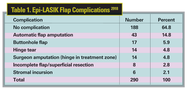
The researchers performed epi-LASIK on 252 eyes of 126 soldiers using the Amadeus II epikeratome and the LADARVision 6000 laser, and compared their results to those from an existing PRK data set collected under identical conditions. The average preop spherical equivalent refraction for the epi-LASIK group was -2.93 ±1.19 D (r: -1 to -6.25 D).
In the epi-LASIK group, 116 cases were available for analysis at three months. There was no significant difference in pain scores between epi-LASIK and PRK at any visit during the first postop week. Re-epithelialization was significantly faster in PRK eyes, however, with 92 percent of them healing by day four, compared to only 55 percent of the epi-LASIK eyes. Ninety-nine percent of the PRK eyes had healed by day seven, compared to 88.5 percent of the epi-LASIK eyes. Fifty epi-LASIK eyes required an additional visit at day 10 postop to confirm complete re-epithelialization, while none of the PRK eyes needed this. Also, epi-LASIK was associated with significantly worse uncorrected acuity at postop day one (p=0.002) compared to PRK. Otherwise, there was no significant difference in uncorrected vision, best-corrected vision or manifest spherical equivalent refraction between the groups through three months postop. The surgeons say it remains to be seen how epi-LASIK compares to PRK in terms of final acuity and haze, but that early healing after PRK is equal or superior to that following epi-LASIK in the first few postop weeks.3354
Surgeons from
Patients with between -4 and -10 D of myopia were randomized to treatment in their dominant eye with either MMC-PRK (0.1 mg/ml for one minute of MMC) or LASEK. The fellow eye underwent conventional PRK. All procedures were performed with the LADARVision 4000 excimer. The researchers measured endothelial cell counts at six and 12 months postop with the Nikon Confoscan.
Postop, 193 patients (97 PRK, 42 MMC-PRK and 54 LASEK) had a follow-up of six months and 138 (69 PRK, 29 MMC-PRK and 40 LASEK) had a follow-up of 12. In the PRK group, there was a median loss of 37 cells/mm^2 (r: -1,092 to 633) at six months and 94 cells/mm^2 (r: -682 to 890) at a year. This change was statistically significant at a year (p=0.001).
For the MMC-PRK group, there was a median loss of 55 cells/mm^2 (r: -574 to 672) at six months and 35 cells/mm^2 (r: -605 to 882) at 12 months, neither of which was statistically significant (p=0.344 and 0.429, respectively).
Additionally, there was no significant change for the LASEK group, with a median loss of 17 cells/mm^2 (r: -936 to 543) at six months and a median gain of 37 cells/mm^2 (r: -789 to 791) at 12 months (p=0.911 and 0.259, respectively). There was a significant difference between the three groups at 12 months (p=0.021); this difference was found to be primarily between the PRK and LASEK groups (p=0.004).3355
Researchers from
In the retrospective study, physicians analyzed the records of 19 eyes with contact lens-intolerant moderate keratoconus (steepest K <60 D; corneal thickness >400 µm) who had undergone PRK with the Zeiss-Meditec MEL-70 or MEL-80 laser using topography-guided ablations.
Postop, best-corrected acuity improved by two or more lines in 14 of the eyes (74 percent). One eye (5 percent) lost two or more lines. After a postop period of six years, one eye needed lamellar keratoplasty.4324
Implant Outcomes
Researchers from ReVision Optics (
The Presbylens is a 1.5-mm diameter hydrogel inlay implanted under a standard LASIK-style flap in a patient's non-dominant eye. The implant is less than 50 µm thick. The researchers say it provides enhanced monovision by creating a multifocal visual pathway in the eye.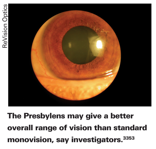
In the study, surgeons implanted the Presbylens in 33 emmetropic presbyopes (mean age: 50 years) whose mean preop spherical equivalent refraction was 0.27 D (r: -0.25 to 0.78) and mean near add was 1.9 D (+1.25 to +2.5 D). Preop, the researchers made sure the subjects could tolerate monovision.
In the treated eyes, uncorrected near vision improved in more than 95 percent of eyes. More than half of the eyes achieved 20/25 or better uncorrected near acuity, compared to zero who saw that well preop. The investigators say that binocular uncorrected distance vision didn't deteriorate significantly postop, and all patients could read the 20/25 line. There were no complications or adverse events.
The researchers also compared the subjects to standard monovision based on their preop defocus blur. Preop, with a 2-D add, all the patients could see 20/20 or better at near, which was superior to the Presbylens' effect. However, the distance vision in the eye with the add would be significantly worse than with the inlay, with none of the eyes seeing 20/25. The researchers say that the inlay significantly improves near vision but doesn't impair distance vision as much as standard monovision.3353
Researchers from the Stanford School of Medicine will present the long-term (three-year) data from the Phase III U.S. Food and Drug Administration investigation of the AMO/Ophtec Artisan (Verisyse) lens for high hyperopia. The trial was sponsored by the lens's maker, Ophtec, and one of the researchers received travel funds from AMO.
In the study, surgeons implanted the lens in 95 eyes of 60 patients at 13 sites. Inclusion criteria included endothelial cell counts of at least 2,000 cells/mm^2 and an anterior chamber depth of at least 3.2 mm. The surgeons followed 46 eyes for three years.
The average implant power used in the study was +7.55 ±2.04 D (r: +4 to +12 D) and the preop anterior chamber depth averaged 3.4 ±0.17 mm. The preop mean spherical equivalent refraction averaged +5.47 ±1.45 D, which was brought down to -0.56 ±0.65 D at year three. Three years after the surgeries, 65.5 percent of eyes were within ±0.5 D of emmetropia, and 98 percent were within ±1 D of it.
Preop, 75.8 percent of eyes saw 20/20 or better best-corrected, with all eyes seeing at least 20/40. Three years after implantation, though, 21 percent could see 20/20 or better uncorrected, and 85.5 percent could see 20/40 or better without correction. In terms of best-corrected acuity postop, 72.7 percent could see 20/20, and all eyes see at least 20/40. Endothelial cell count decreased by an average of 4.3 percent over three years, from 2,588 to 2,400 cells/mm^2 (p<0.01).5646
Ablation Efficiency
Researchers from
In the study, the investigators used a Technolas 217 (Bausch & Lomb), a LADARVision 4000 and an Allegretto Wave Eye-Q laser to ablate both flat and spherical artificial eyes made of filofocon A, a fluoro-silicon-acrylate material. They applied myopic corrections of 3, 6 and 9 D, using optical zones of 6.5 mm. In-eye artificial pupils allowed centration similar to patients' eyes.
The researchers measured the ablation shapes with a precision of 1 µm, both on flat and spherical surfaces. Ablation depths were 0.34 (Technolas), 0.54 (LADARVision) and 0.44 (Allegretto) times the nominal value on the cornea. The postop apical radius of curvature varied across lasers: For the 9-D correction, for example, the Technolas radius of curvature was 8.12 ±0.11 mm; the LADAR resulted in 8.52 ±0.05 mm and the Allegretto resulted in 7.93 ±0.06 mm. The induced asphericity also varied: 0.28 ±0.11 for the Technolas; 0.26 ±0.10 for the LADAR and -1.6 ±0.10 for the Allegretto. This induced asphericity resulted in different amounts of spherical aberration in the identical artificial eyes.
The researchers say that they found the eye models to be useful in assessing the effects of different lasers or ablation algorithms, calibrating a laser, and calculating a correction factor to improve postop corneal shape.2445
Dr. Probst is medical director for TLC Laser Centers.