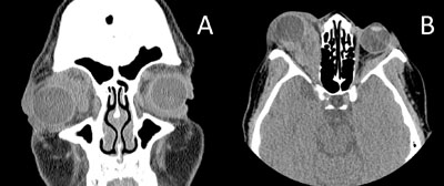
Laboratory blood work was sent, including TSH, T3, free T4 and thyroid stimulating immunoglobulin, which all came back within normal limits. The patient underwent further imaging to characterize the orbit pathology. Computed tomography of the orbits without contrast (See Figure 2) was initially performed and showed extensive soft tissue infiltration throughout the right orbit, including near-complete involvement of the retrobulbar fat and obscuration of the optic nerve intraconally. Proptosis and mild tenting of the posterior right globe surface was noted. The right medial rectus muscle was markedly enlarged, including thickening of its tendinous insertion, while the remaining EOMs were normal in appearance. Additionally, there was abnormal enlargement of the left lateral rectus muscle. The orbital walls were intact. Given these findings, thyroid-related disease was considered less likely due to tendon involvement and the atypical pattern of EOMs affected.
Upon further questioning, it was ascertained that the patient had noticed mild nipple discharge from both breasts for a couple of months and never had a prior mammogram. A breast exam was subsequently performed which revealed an extensive right breast mass with overlying inflammatory skin changes and spread across the mediastinum toward the contralateral breast. Rapid progression of proptosis and possible early optic neuropathy warranted admission of the patient to expedite management and an orbitotomy with biopsy of the orbital lesion was performed. The final pathology report indicated morphologic and immunohistochemical findings consistent with breast carcinoma; furthermore, the specimen was estrogen receptor (ER) and progesterone receptor (PR) positive but human epidermal growth factor receptor (Her2) negative. Tumor markers were also sent and revealed normal CEA and CA-125; however, CA 15.3 was elevated at 100 (reference range <25).
|
Discussion
Breast carcinoma most frequently metastasizes to liver, bone, lungs, skin and brain.1 Only a small percentage of cases are associated with disease spread to the orbit, with one study showing an overall rate of 0.2 percent.2 However, breast carcinoma is considered to be the most prevalent primary tumor of all metastatic tumors to the orbit, with an estimated prevalence ranging from 28.5 to 58.8 percent of all orbital metastases.3 In up to 25 percent of cases, orbital metastasis is the initial finding of a previously undetected primary malignancy.4
The presentation of orbital metastasis is mostly unilateral but can affect right and left sides equally. The anatomical distribution within the orbit predominantly involves the superior and lateral quadrants. Various ocular signs and symptoms are commonly reported, including proptosis, eyelid swelling or mass, pain, ptosis, bulb divergence and blurred vision. Diplopia resulting from motility deficits is a prevalent symptom and occurs as a result of tissue-specific preference of breast cancer to extraocular muscles and surrounding orbital fat. Separately, enophthalmos is a less common but characteristic sign of orbital infiltration by the serous subtype of breast adenocarcinoma.4,5
|
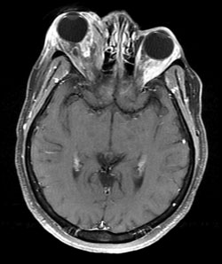
For evaluation of orbital masses, magnetic resonance imaging with gadolinium contrast is considered the imaging method of choice. MRI is advantageous over computerized tomography because of higher soft tissue contrast and lack of radiation exposure.9 Orbital metastases appear on non-contrast CT as irregularly shaped lesions that are isodense to muscle and demonstrate slight enhancement with contrast. Orbital bony wall involvement is also a common finding. On MRI, metastatic disease is hypointense to fat on T1-weighted images and hyperintense to fat on T2-weighted images. This is characteristically distinct from orbital pseudotumor, which is isointense to fat on T2WI.4
Overall, orbital surgery geared toward removal of metastasis is not recommended because the procedure is not curative and may incur significant ocular morbidity.10 Likewise, other radical surgeries or enucleation do not offer benefit in terms of survival and are only considered in cases complicated by intractable ocular pain or uncomfortable symptoms related to rapid tumor growth. At this time, the only surgical intervention recommended for breast carcinoma that has metastasized to the orbit is a tissue biopsy (either FNA or open biopsy) to establish the diagnosis.11
Findings of orbital extension from a primary breast carcinoma predict widespread disease to other organs.1 The median survival time for patients with orbital metastasis of breast malignancy is reported to be 22 to 31 months.4 Thus, the mainstay treatment is palliative radiotherapy, with clinical improvement of symptoms and vision in 60 to 80 percent. External-beam irradiation is the most common modality.11,12 Two alternative modalities include stereotactic radiation therapy and stereotactic radiosurgery, both of which may allow better quality of life due to the application of high doses of radiation to a well-defined volume and shorter treatment courses. Pending performance status, chemotherapy and/or hormone therapy in patients harboring hormone-sensitive tumors may be indicated.13,14
As for post-chemotherapy or post-radical treatment surveillance, several serum-based products and tumor markers are utilized in the management of breast cancer patients. Of these biomarkers, CA 15.3 has been extensively evaluated in the literature. A 2012 study concluded that CA 15.3 is one of the most powerful tools for early detection of breast cancer recurrence, as well as a cost-reducing tool for chemotherapy monitoring.15
Our case demonstrates that when presented with an infiltrative orbital process, the differential diagnosis of breast cancer must be considered, especially in women. Despite the availability of sophisticated imaging modalities, obtaining a careful history and having a low threshold for diagnostic breast examination is recommended to aid diagnostic efforts.
The author would like to thank Nicolas Biro, MD, of the Neuro-Ophthalmology Service for his time and assistance in preparing this case report.
1. Francone E, Murelli F, Paroldi A, Margarino C, Friedman D. Orbital swelling as a first symptom in breast carcinoma diagnosis: A case report. J Med Case Rep 2010 Jul 15;4:211.
2. Tamura M, Tada T, Tsuji H, et al. Clinical study on the metastasis to the eyes from breast cancer. Breast Cancer 2004;11: 65-8.
3. Eckardt AM, Rana M, Essig H, Gellrich NC. Orbital metastases as first sign of metastatic spread in breast cancer: Case report and review of the literature. Head Neck Oncol 2011;3:37.
4. Vlachostergios PJ, Voutsadakis IA, Papandreou CN. Orbital metastasis of breast carcinoma. Breast Cancer (Auckl). 2009;3:91-7.
5. Gupta S, Bhatt VR, Varma S. Unilateral orbital pain and eyelid swelling in a 46-year-old woman: Orbital metastasis of occult invasive lobular carcinoma of breast masquerading orbital pseudotumour. BMJ Case Rep 2011; Published online 2011 March 15.
6. Reeves D, Levine MR, Lash R. Nonpalpable breast carcinoma presenting as orbital infiltration: Case presentation and literature review. Ophthal Plast Reconstr Surg 2002;18:84-8.
7. González F, López-Couto C. Orbital metastases. A report of four cases and a review of the literature. Arch Soc Esp Oftalmol 2006;81:451-62.
8. Shields JA, Shields CL, Scartozzi R. Survey of 1,264 patients with orbital tumors and simulating lesions: The 2002 Montgomery Lecture, part 1.Ophthalmology. 2004;111:997-1008.
9. Tomizawa Y, Ocque R, Ohori NP. Orbital metastasis as the initial presentation of invasive lobular carcinoma of breast. Intern Med 2012;51(12):1635-8.
10. Char DH, Miller T, Kroll S. Orbital metastases: diagnosis and course. Br J Ophthalmol 1997;81:386-390.
11. Mehdi AS, Esmaeli B. Metastatic tumors of the orbit and ocular adnexa. Curr Opin Ophthalmol 2007;18:405-413.
12. Shields JA, Shields CL, Brotman HK, et al. Cancer metastatic to the orbit: The 2000 Robert M. Curtis Lecture. Ophthal Plast Reconstr Surg 2001;17:346-54.
13. Bellmann C, Fuss M, Holz FG, et al. Stereotactic radiation therapy for malignant choroidal tumors. Ophthalmology 2000;107:358-65.
14. Kouvaris JR, Gkongkou PV, Papadimitriou CA, et al. Bilateral metastases to extraocular muscles from lobular breast carcinoma. Onkologie 2008;31:387-9.
15. Bahrami-Ahmadi A, Makarian F, Mortazavizadeh MR, et al. Symptomatic metastasis prediction with serial measurements of CA 15.3 in primary breast cancer patients. J Res Med Sci 2012;17(9):850-4.


