Background
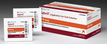
Gamma-aminobutyric acid is the brain’s major inhibitory neurotransmitter. Vigabatrin (Sabril) is a selective, irreversible, inhibitor of GABA transaminase that increases levels of GABA in the brain; it was synthesized in the 1980s and 1990s as an anti-epileptic medication.1,2 The molecule resembles GABA, but the addition of an extra vinyl group allows it to sit in the active site of GABA transaminase and render it inactive. It has been used around the world since the 1990s and was recently approved for use in the United States. Vigabatrin is often effective for the management of refractory complex partial seizures in adults who have failed multiple medications. Such patients are often debilitated by their seizures and are desperate for treatment. Compared with other anti-epileptics, vigabatrin has a relatively low interaction rate with other medications, a lower overall rate of side effects, and is particularly effective when spasms are associated with tuberous sclerosis.3,4 In children, it is an option for first-line therapy for infantile spasms (West syndrome), which are seizures hypothesized to be malfunctions of the GABA regulation process and are notoriously difficult to control. Adrenocorticotropic hormone (ACTH) can also be used as first-line treatment for infantile spasms, since excess production of corticotropin-releasing hormone is another possible etiology, but this treatment carries a higher risk profile.
In 1997, Dr. Tom Eke and colleagues described cases of peripheral visual field constriction associated with vigabatrin.5 Since then, vigabatrin has clearly been shown to cause a dose-dependent, permanent peripheral field constriction.2,6 Other, less common side effects include somnolence, headache, dizziness, fatigue and weight gain. Psychosis has also been reported, although this side effect is more common in adults than in children. The prevalence of visual field constriction is uncertain and ranges from 14 to 92 percent in various studies. Because of this side effect, nearly two decades of debate delayed the approval of vigabatrin in the United States until August 21, 2009. Prior to approval, patients and their family had to obtain vigabatrin from Canada, Mexico and the United Kingdom. After approval, the Food and Drug Administration implemented a Risk Evaluation and Mitigation Strategy (REMS) to promote compliance with its screening recommendations. The earliest reports of toxicity were after 11 months of exposure. The vision loss is usually asymptomatic and spares the macula, but sub-clinical depression of macular function and color vision deficits have been reported.
 |
Current recommendations limit the dose of vigabatrin to 3 g per day in adults, or 50 to 100 mg/kg/day in children, and the drug should be withdrawn if it does not provide adequate seizure control.9 Adult ophthalmologists will be seeing patients on vigabatrin more frequently as its uses expand. Robert Fechtner, MD, and colleagues recently showed that vigabatrin is effective in treating cocaine and methamphetamine dependence.10 Their study enrolled 28 patients and found that 16 remained negative for substance use during the last six weeks of the study. Furthermore, no ocular adverse effects were reported.
Use of vigabatrin involves a continuous analysis of its risks and benefits. This approach requires cooperation among the patient’s neurologist, ophthalmologist and family. Some families will accept risk of a visual field defect if vigabatrin provides freedom from seizures, while others would prefer not to risk visual impairment.
Toxicity Evaluation Options
The FDA has recommended that all patients have complete eye examinations and visual field testing before starting vigabatrin. Patients should return for follow-up exams every three months to monitor for side effects. Unfortunately, detection of visual field defects in this population of children is very difficult. The majority of children who require vigabatrin for seizure control are very young, non-verbal, or are unable to cooperate with the most sensitive tests. The American Academy of Pediatric Ophthalmology & Strabismus has recommended alternative ways to evaluate for toxicity:
• Serial fundus examinations. The appearance of the fundus may be completely normal despite toxicity and visual field constriction; however, indirect ophthalmoscopy may be the best method for a large proportion of pediatric patients. Optic nerve findings include thinning of the nasal retinal nerve fiber layer often referred to as “reverse optic atrophy.”11-13 Macular pigment epithelial changes have also been described.14-16
• Serial automated static perimetry. Reliable results are usually only achievable in older children, at least 9 years old, who are able to cooperate for the examination. Formal visual field testing performed on younger children, even those who can sit for the test, is not reliable or sensitive enough to detect early subtle changes. Recent studies also suggest that perimetry alone may not be enough to prove vigabatrin toxicity. Dr. Gonzalez and coworkers illustrated bilateral visual field constriction in 24 percent of vigabatrin-naive epileptic patients and concluded that visual fields constriction alone is not necessarily indicative of medication toxicity.8
• Optical coherence tomography. OCT has revolutionized our understanding of many different diseases that affect the nerve fiber layer and is a useful tool for detection of nerve fiber layer thinning in adults and older, cooperative children. Lisa Clayton and colleagues recently illustrated the effectiveness of OCT for evaluation of patients taking vigabatrin.17 In this study, the average retinal nerve fiber layer thickness in patients taking vigabatrin was significantly thinner than in healthy controls. The extent of the nerve fiber layer thinning correlated well with the extent of visual field loss. The authors conclude that OCT is a reliable and objective tool for evaluating patients on vigabatrin. Unfortunately, it will not provide reliable information on younger children and patients who are unable to cooperate. Recent development of a handheld supine OCT may improve screening for vigabatrin toxicity in infants and young children. Such devices have been used successfully in a subset of these patients.
• Visual evoked potentials. Graham Harding, DSc, and colleagues illustrated that VEP can be used to detect visual field defects secondary to vigabatrin.18 Their field-specific stimulus identified three of four abnormal perimetry results and seven of eight normal perimetry results, giving a sensitivity of 75 percent and a specificity of 87.5 percent. The authors conclude that field-specific VEPs are well-tolerated by children older than 2 years and are sensitive and specific enough to identify vigabatrin-associated defects. Unfortunately, despite the results of such studies, the authors conclude that “ …in individual subjects, the tests are simply too unreliable to guide decision-making with regards to vigabatrin maintenance.”9
• Electroretinograms. ERGs have been shown to detect early changes associated with vigabatrin toxicity; however, several problems prevent ERG from being an ideal screening tool. A recent report warned against over-reliance on the ERG to detect vigabatrin toxicity. Their study found no significant association of any ERG parameter with visual field defects and could not determine if the ERG abnormalities they found were due solely to the effects of vigabatrin. Furthermore, there is no accepted “normal” waveform for very young children, and ERG in the pediatric population often requires general anesthesia, which may also alter the waveform. Finally, general anesthesia every three months is not convenient and is potentially dangerous. Most parents are hesitant to accept subjecting their child to the risks inherent in general anesthesia.
In conclusion, vigabatrin is a very effective drug for treatment of infantile spasms and seizure disorders refractory to other medications. Its use, however, is complicated by an irreversible, dose-dependent visual field constriction from photoreceptor toxicity. The majority of patients are asymptomatic since the macula is usually spared. Screening for this side effect in young children can be very difficult, especially given the fact that many children who need vigabatrin are non-verbal and poorly cooperative. Methods of screening for visual field constriction include serial fundus examination, serial automated static perimetry, OCT, VEP and ERG. Each method has its advantages and disadvantages. Most commonly, parents opt for serial fundus examination and prefer to avoid repetitive general anesthesia for their child. “Reverse” optic atrophy and macular pigment epithelial changes are the most common ophthalmoscopic findings. Any ophthalmologist screening children on vigabatrin should be ready to discuss the options with parents so they can make informed decisions. In addition, a periodic re-evaluation of the need for vigabatrin should be initiated by the patient’s neurologist to ensure patients do not needlessly remain exposed to the risk of vision loss. The AAPOS policy statement on vigabatrin can be found at: http://www.aapos.org//client_data/files/2012/504_vigabatrin_05.09.12.pdf. Hopefully, future research will discover more effective tests of peripheral vision for patients who cannot comply with traditional methods. REVIEW
Dr. Fecarotta is an assistant clinical professor of ophthalmology at SUNY Downstate Medical Center in Brooklyn, N.Y.
1. Dichter MA, Brodie MJ. New antiepileptic drugs. N Engl J Med 1996;334:1583-90.
2. Ben-Menachem, E. 1995 Vigabatrin. Epilepsia 36 (Suppl. 2), S95-S104.
3. Camposano S, Major P, Halpern E, Thiele E. Vigabatrin in the treatment of childhood epilepsy: A retrospective chart review of efficacy and safety profile. Epilepsia 2008;49:1186-91.
4. Hancock E, Osborne J. Vigabatrin in the treatment of infantile spasms in tuberous sclerosis: Literature review. J Child Neurol 1999;14:71-74.
5. Eke T, Talbot JF, Lawden MC. Severe persistent visual field constriction associated with vigabatrin. BMJ 1997;314:180-1.
6. Bruni J, Guberman A, Vachon L, Desforges C. Vigabatrin as add on therapy for adult complex partial seizures: A double blind, placebo-controlled multicentre study. The Canadian Vigabatrin Study Group. Seizure 2000;9:224-232.
7. Jammoul F, Wang Q, Nabbout R, et al. Taurine deficiency is a cause of vigabatrin-induced retinal phototoxicity. Ann Neurol 2009 Jan: 65(1):98-107.
8. Gonzalez P, Sills GJ, Parks S. Binasal visual field defects are not specific to vigabatrin. Epilepsy Behav 2009 Nov;16(3):521-6. doi: 10.1016/j.yebeh.2009.09.003. Epub 2009 Oct 7.
9. http://www.aapos.org//client_data/files/2012/504_vigabatrin_05.09.12.pdf.
10. Fechtner RD, Khouri AS, Figureoa E, Ramirez M, et al. Short term treatment of cocaine and/or methamphetamine abuse with vigabatrin: Ocular safety pilot results. Arch Ophthalmol 2006;124:1257-62.
11. Frisen L, Malmgren K. Characterization of vigabatrin-associated optic atrophy. Acta Ophthalmol Scand 2003;81:466-73.
12. Wild JM, Robson CR, Jones AL, Cunliffe IA, Smith PE. Detecting vigabatrin toxicity by imaging of the retinal nerve fiber layer. Invest Ophthalmol Vis Sci 2006;47:917-24.
13. Buncic JR, Westall CA, Panton CM, Munn JR, McKeen LD, Logan WJ. Ophthalmology 2004;101:576-580.
14. French JA, Mosier M, Walker S, Sommerville K, Sussman N. A double-blind, placebo-controlled study of vigabatrin three g/day in patients with uncontrolled complex partial seizures. Vigabatrin Protocol 024 Investigative Cohort. Neurology 1996;46:54-61.
15. Guberman A, Bruni J. Long-term open multicentre, add-on trial of vigabatrin in adults resistant partial epilepsy. The Canadian Vigabatrin Study Group. Seizure 2000;9:112-118.
16. Sander JW, Trevisol-Bittencourt PC, Hart YM, Shorvon SD. Evaluation of vigabatrin as an add-on drug in the management of severe epilepsy. J Neurol Neurosurg Psychiatry 1990;53: 1008-1010.
17. Clayton LM, Devile M, Punte T, et al. Retinal nerve fiber layer thickness in vigabatrin- exposed patients. Ann Neurol 2011;69:845-854.
18. Harding GFA, Spencer EL, Wild JM, Conway M, Bohn RL. Field-specific visual evoked potentials: Identifying field defects in vigabatrin-treated children. Neurology 2002; 58(8):1261-1265.


