Mitomycin's Efficacy
As explained by Rajiv Mohan, PhD, of the Cleveland Clinic, mitomycin is an alkylating agent that forms a covalent linkage with DNA and inhibits DNA synthesis. "Higher concentrations block cellular RNA and protein synthesis," he explains. "This is how it affects the keratocyte density in the cornea, which, in turn, affects corneal haze. The keratocyte is a very slow, non-dividing cell. When you do PRK, it triggers a wound-healing response in which cytokines are released from the corneal epithelium, activating the transformation of keratocytes into myofibroblasts, which are actively dividing cells that are sometimes called 'activated keratocytes.' Active division causes the cells to lose their crystalline formation and structure, making them hazy. As an alkylating agent, mitomycin blocks the DNA synthesis, inhibiting the proliferation and, therefore, the haze formation."
Many surgeons use mitomycin successfully with their surface procedures, and some of them have published their results as proof of the agent's efficacy.
In one prospective study from Italy, surgeons divided 62 highly myopic (greater than -5 D) PRK patients (124 eyes) into two equal groups. One group received a prophylactic mitomycin treatment during their surgeries, while the other did not.
Though mitomycin has been an effective haze preventer for many, a 0.04%, 50-second application didn't prevent haze from occurring in this hyperopic LASEK eye.
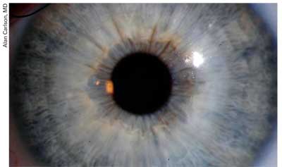
Alan Carlson, MD
At one year postop, the best-corrected vision of the mitomycin group was better than that of the control group (p=0.013). In the mitomycin group, 43 eyes (69 percent) were within 0.50 D of attempted correction, compared to 31 eyes (50 percent) of the control group. There was also a smaller incidence of corneal haze in the mitomycin group (p=0.005).1
Another study that was prospective, masked and randomized divided 60 consecutive eyes of 60 patients into two groups: one that received a two-minute application of 0.2 mg/ml after ablation; and a control group that didn't receive mitomycin.
The surgeons reported no toxic side effects, and comment that no mitomycin eye had a haze rate greater than grade 1 during the six-month follow-up, compared to 19 eyes (63 percent) in the control group (p=0.01). At six months, the difference in refractive outcome between the groups was statistically significant (p=0.05), with 26 study-group eyes (87 percent) and 14 controls (47 percent) within ±0.50 D of the attempted correction. No mitomycin eye lost best-corrected acuity during follow-up, while seven control eyes lost one to three lines at six months (p=0.0006).2
Surgeons have also reported using it safely in conjunction with transepithelial PTK/PRK over complicated LASIK flaps. In a study of 10 such patients, no one experienced delayed re-epithelialization, haze or other signs of toxicity.3
Mitomycin Toxicity: What We Know
Despite such successes, some surgeons think it might be worth taking a step back and evaluating the use of mitomycin to ensure they and their patients don't get any nasty surprises in the future.
Dr. Mohan thinks mitomycin has the potential for problems if it's not used solely on selected patients and under particular protocols for concentration and duration.
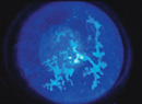
Rabbits treated with 0.02% mitomycin for two minutes (right) demonstrated a central zone devoid of keratocytes (the black area devoid of cellular specks) six months postop, while animals who received BSS (left) did not.

Rajiv Mohan, MD
"Surgeons are just using mitomycin left and right, but there have been no systematic studies performed," Dr. Mohan says. "We did see what mitomycin can do, though, at least in the rabbit."
In Dr. Mohan's study, published in the Journal of Cataract and Refractive Surgery, researchers performed -9 D PRK ablations with either mitomycin or BSS on 224 rabbits.4 They used mitomycin concentrations of 0.02% or 0.002%, and three different durations of application: 12 seconds; one minute and two minutes.
"Essentially, the results were very similar in inhibiting the haze, but the tissue that was exposed to 0.02% mitomycin for two minutes had significantly reduced keratocyte density, even after six months," says Dr. Mohan. "In fact, even at six months we saw an acellular zone in the anterior stroma where the PRK had been performed. This area was devoid of all keratocytes, which raises a large red flag. Such a reduction of keratocyte density in the anterior stroma may cause biomechanical instability, iatrogenic ectasia and possibly corneal melting. It's possible that, after a longer follow-up, the keratocytes will repopulate. However, this is unlikely because six months is a long time."
Dr. Mohan says that, given what his group found, he and his colleagues would recommend that surgeons use the lower concentration and duration, since the researchers didn't see such a depletion of keratocytes in the lower dose/shorter exposure groups, though the haze inhibition was relatively the same. "Also, we recommend using mitomycin mainly for higher dioptric corrections, starting with a minimum of -5 or -6 D, rather than for every patient undergoing PRK," he adds.
Though a rabbit's cornea isn't exactly the same as a human's, Dr. Mohan thinks the results can still help as a guideline, based on earlier studies he performed that examined similarities between the two.
"When I was at the University of Washington, Seattle, we scraped the epithelium in human eyes that were due to be removed due to cancers or other reasons, and compared the corneas to rabbit corneas that we had scraped," he explains. "We performed TUNEL assays and other tests, and found cellular reactions similar to those we saw in the rabbit."
Duke University corneal specialist Alan Carlson echoes Dr. Mohan's sentiments, saying that the dose/response curve of mitomycin needs defining. "To me, the most significant concern with mitomycin relates to our lack of a defined therapeutic window guiding our treatment, both in terms of concentration and duration of application," he says. "To this point, it's been trial and error. We know that using too much of it risks significant toxicity, cell loss, poor healing and scarring. As we lower the dosage, however, people are clinically reporting that it works, even when we get down to what we might consider homeopathic doses. We don't know."
A significant study that was recently published in the American Journal of Ophthalmology may give refractive surgeons pause as they consider the use of mitomycin.5
In the prospective, randomized, double-blind, placebo-controlled study, nine patients received a prophylactic mitomycin application at the time of their PRK in one eye and BSS in the fellow eye. The application was performed after the ablation, using 0.02% mitomycin for 30 seconds. The surgeons performed corneal pachymetry and endothelial cell counts at one month and three months, and compared the results to measurements taken preoperatively.
Preop, there was no significant difference in the endothelial cell count between the treated and control eyes. The mitomycin group's count was 2,835 ±395, the control group's was 2,779 ±492 (p=0.62). At one month and three months postop, however, the endothelial cell loss was statistically significant in the mitomycin group: The loss at one month was 14.7 ±5.1 percent; and at three months it was 18.2 ±9 percent (p=0.0006 and p=0.002, respectively). In the control eyes, at one month and three months the difference in the endothelial cell count wasn't statistically significant (p=0.27 and p=0.14, respectively).
"Those are significant numbers when compared to the normal range," muses Dimitri Azar, MD, corneal specialist and head of the department of ophthalmology and visual science at the University of Illinois' Eye and Ear Infirmary in Chicago. "In light of them, when I speak to my patients about mitomycin I will now note that the number-one side effect is decrease of endothelial cell counts, based on this report." He says that he might not use it prophylactically now in someone with very low endothelial cell counts, but would still use it therapeutically postop for a patient who had developed haze and needed treatment. He also takes some comfort in that the study's dose was 10 times stronger than the one he normally uses prophylactically, and was used for twice his usual amount of application time.
Roswell Pfister, MD, of Eye Research Laboratories in Birmingham, Ala., also thinks the numbers are significant, but would be interested to see if the trend continues over time.
"No one really knows what the agent's long-term effects will be," he says. "Recall one thing though: The endothelial cell count progressively decreases as a person ages. The question is, will the numbers found in this study continue to go down over time? Three months follow-up isn't very long."
Dr. Pfister himself reported on one of the only other cases of endothelial cell loss after mitomycin use on the cornea that appears in the literature in a 2004 article in Cornea.6 In his case report, a 39-year-old man underwent PTK for recurrent corneal erosions secondary to basement membrane dystrophy, and subsequently developed irregular astigmatism and central stromal opacity. The cornea was scraped and treated with 0.02% mitomycin drops over a period of six days, receiving 14 drops total. The patient eventually developed corneal edema due to a low endothelial cell count and dysfunctional cells, and went on to have a corneal transplant.
In the study, Dr. Pfister makes a point to say he feels a one-time mitomycin application at the end of a surgery is a valuable adjunct when no other option is available, and is different from a prolonged topical application of the drug such as the study patient had undergone. He offers the case merely so surgeons will be aware of a potential pitfall of the agent if and when they use it.
It's in the Way That You Use It
Surgeons say that, though mitomycin may pose a risk for a rare complication such as endothelial dysfunction, the judicious use of it can go a long way toward ensuring that these complications remain uncommon.
"We have been very enthusiastic about mitomycin as an agent to treat corneal haze, as opposed to a way to prevent haze formation," says Dr. Azar. "We have done studies looking at its use in rabbit eyes, published several years ago just after the first human reports came out, that confirmed that it's an effective way to treat corneal haze and scar formation after PRK. But, at the same time, given the potential side effects that are known to ophthalmologists with regard to the use of mitomycin after glaucoma filtering surgery, for ocular surface tumors and especially for pterygium removal, one has to be careful before recommending the treatment as a prophylactic approach. To use it prophylactically involves applying the drops to many, many more patients than its therapeutic use does, because less than 3 percent of patients under 6 D of myopia will develop haze or scarring. So, 97 percent will be getting an application without really benefiting from it." As a therapeutic use for someone who's developed haze, Dr. Azar uses 0.02% for 30 seconds and has been very pleased with the results.
For prospective PRK patients who may benefit from a prophylactic application, however, Dr. Azar proceeds carefully and with an in-depth discussion with the patient, taking several risk factors for haze into account. He considers where the patient lives and his skin pigmentation—factors which may play a role in haze formation—the degree of myopia, and now, probably his endothelial cell count.
"Other factors can be the degree of allergy and/or dry eye, as well as what medications the patient in taking," says Dr. Azar. "So, if you can characterize a patient as being at high risk for haze due to these factors, it may make sense to discuss the possibility of using mitomycin prophylactically. If you can't, then it may be overkill to use the agent." For a prophylactic treatment, Dr. Azar uses 0.002% for 12 seconds.
Wills Eye Hospital corneal specialist Christopher Rapuano also thinks it's probably "overkill" to use mitomycin prophylactically on all PRK patients. "I use it in the higher myopic patients, usually above 5 or 6 D," he says. "I use the 0.02% concentration for 12 seconds, applying it with a circular, 6.5 mm cellulose sponge, making sure it's on the sponge and doesn't go all over the eye, such as onto the conjunctiva. When I'm done, I dry the sponge before I lift it from the eye, to avoid leaving any excess. I then irrigate the eye with 30 cc of saline."
In an effort to perform the safest application possible, Dr. Azar uses a Merocel sponge fashioned into a ring, rather than the disk that's used by some surgeons.
"I put two punches in a 1-mm thick Merocel sponge, creating a ring with a 3.5 mm inside diameter and a 5.5 mm outside diameter," he explains. "By using a disk, the medicine goes in but the concentration in the center of the cornea is very low on the deeper levels. In this way, if there's any endothelial toxicity, it's just in the small area on which the ring was applied, rather than the whole cornea. The reason it works is because the fibroblasts will migrate from the periphery to the center, so it will limit their ability to divide but their migration won't be affected.

Chicago surgeon Dimitri Azar applies mitomycin using a ring in order to spare the center from direct exposure.
"If a surgeon doesn't have the materials available to make the ring, he can punch out a small Merocel sponge, soak it with the mitomycin, then move it in a circular fashion around the outer part of the treatment zone. He then irrigates the eye as usual."
Some surgeons may consider reducing their duration of application even further, as studies such as Dr. Mohan's emerge to report that short durations don't seem to negate the efficacy, yet may decrease the risk of a complication.
"I have generally reserved it for myopes in the 10 D-to-12 D range for about two minutes," says Dr. Pfister. "And I've had favorable outcomes. But one article [Dr. Mohan's] reported reducing the time from two minutes, to one, then down to 12 seconds and found similar effects from the various durations. Even the glaucoma surgeons who are using it for trabeculectomy have reduced their application time over the years; they used to do it for five minutes, now some are going down to one minute to avoid the possibility of an excessive effect and a hypotonous eye postop."
Dr. Mohan's recommended protocol for prophylactic mitomycin is exactly like Dr. Azar's, except for Dr. Azar's addition of the ring rather than the disk. "Based on our rabbit study, you can use 0.002% mitomycin for 12 seconds," says Dr. Mohan. "This is very effective, and you'll see a very minor difference in the level of decreased haze. But if you compare the risk vs. benefit, I think this may be much safer for the patient long term."
As surgeons continue to use mitomycin judiciously, time will reveal whether the protocols are safe or need further modifications to ensure safety.
"Five years from now, are we going to be using mitomycin the same way we're using it now?" wonders Dr. Carlson. "I hope not. But you can say the same thing about cars, computers and other things. I think now we're doing a pretty good job of safely selecting patients for laser surgery, but in five years I think we're going to have better lasers, flaps and ways of doing things, and we're going to think of things that we wish we knew when we were using mitomycin in the manner in which we do now."
1. Bedei A, Marabotti A, Giannecchini I, et al. Photorefractive keratectomy in high myopic defects with or without intraoperative mitomycin C: 1-year results. Eur J Ophthalmol 2006;16:2:229-34.
2. Carones F, Vigo L, Scandola E, Vacchini L. Evaluation of the prophylactic use of mitomycin-C to inhibit haze formation after photorefractive keratectomy. J Cataract Refract Surg 2002;28:12:2088.
3. Muller LT, Candal EM, Epstein RJ, Dennis RF, Majmudar PA. Transepithelial phototherapeutic keratectomy/photorefractive keratectomy with adjunctive mitomycin-C for complicated LASIK flaps. J Cataract Refract Surg 2005;31:2:291-96.
4. Netto MV, Mohan RR, Sinha S, et al. Effect of prophylactic and therapeutic mitomycin C on corneal apoptosis, cellular proliferation, haze, and long-term keratocyte density in rabbits. J Refract Surg 2006;22:6:562-74.
5. Morales AJ, Zadok D, Mora-Retana R, Martinez-Gama E, Robledo NE, Chayet AS. Intraoperative mitomycin and corneal endothelium after photorefractive keratectomy. Am J Ophthalmol 2006;142:3:400-4.
6. Pfister RR. Permanent corneal edema resulting from the treatment of PTK corneal haze with mitomycin: A case report. Cornea 2004;23:7:744-47.