 |
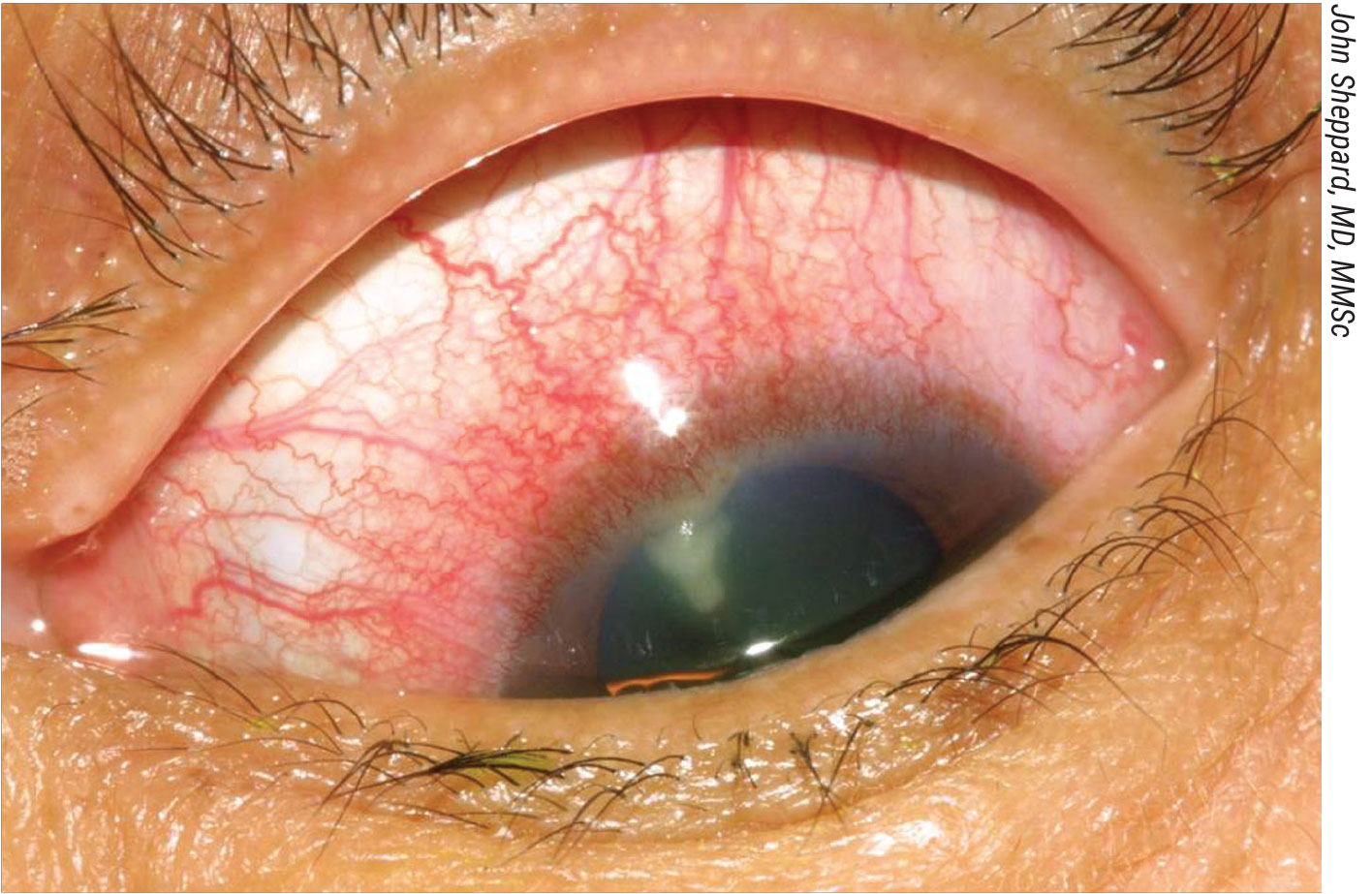
| Staphylococcal infections demonstrated greater susceptibility to even some older medications in the newest ARMOR study. |
Despite encouraging positive trends for S. aureus, multidrug resistance—microbial insensitivity to at least three medication classes—is still common for many organisms.
Treating ocular infections is hard enough as is when the drugs work as advertised, and so much the worse when the offending microorganism is resistant or only weakly susceptible to therapy. Staphylococci are known causative pathogens in ophthalmic infections, and antibiotic resistance among these bacteria is of clinical concern. The long-running Antibiotic Resistance Monitoring in Ocular micRoorganisms (ARMOR) Study, the only nationwide surveillance study of its kind, captures in vitro data specific to common ocular pathogens. With two research posters, the same team presented their findings on the 14th year of the study’s data collection at ARVO 2023 in New Orleans. Each noted that, with preliminary data indicating lower resistance rates especially among Staphylococcus aureus, multidrug resistance was common among methicillin-resistant strains.
One analysis reported on 2022’s data, when 397 isolates were collected January through October of that year.1 Staphylococcus aureus, coagulase-negative staphylococci (CoNS), Streptococcus pneumoniae, Pseudomonas aeruginosa and Haemophilus influenzae from ocular infections were collected as part of ARMOR and submitted to a central laboratory for species confirmation and in vitro antibiotic susceptibility testing. Minimum inhibitory concentrations for up to 16 antibiotics (10 drug classes) were determined and interpreted.
The 142 CoNS isolates exhibited the highest resistance, with azithromycin, oxacillin/methicillin, trimethoprim, clindamycin and tetracycline resistance observed in 60 percent, 37 percent, 28 percent, 27 percent and 22 percent of isolates, respectively. Among the 161 S. aureus isolates, 46 percent were resistant to azithromycin, but <20 percent of isolates were resistant to other drugs. Multidrug resistance (poor or ineffective response to three or more drug classes) was observed in 14 percent of S. aureus, 39 percent of CoNS and in 59 percent and 88 percent of methicillin-resistant strains thereof, respectively. Among the five S. pneumoniae isolates, 60 percent were resistant to azithromycin, oral penicillin and tetracycline. Although all 72 P. aeruginosa isolates were resistant to polymyxin B, <5 percent were resistant to other drugs; no resistance was found among the 17 H. influenzae isolates.1
“The clinical significance of these in vitro data is unclear without consideration of the ocular pharmacokinetics of tested antibiotics,” the ARMOR researchers concluded in their abstract.1
The team’s other study examined resistance trends over time among staphylococcal isolates collected from 2009 through 2022 in the ARMOR study. A total of 2,999 S. aureus and 2,575 CoNS were included in their analysis.2
In vitro resistance decreased to methicillin/oxacillin (S. aureus, 39 percent in 2009 to 18 percent in 2022; CoNS, 50 percent in 2009 to 37 percent in 2022) and to ciprofloxacin (S. aureus, 39 percent in 2009 to 17 percent in 2022; CoNS, 46 percent in 2009 to 20 percent in 2022). Additionally, among S. aureus, resistance to azithromycin decreased (62 percent in 2009 to 46 percent and 9 percent in 2022), as did resistance to tobramycin (24 percent in 2009 to 9 percent in 2022), while in contrast an increase in chloramphenicol resistance was observed (7 percent in 2009 to 3 percent in 2022, peaking at 30 percent in 2021). Cumulative multidrug resistance (three or more antibiotic classes) was observed in 30 percent of S. aureus and 41 percent of CoNS and in 76 percent and 79 percent of methicillin-resistant isolates thereof, respectively.
“ARMOR continues to inform us about ocular infections and antibiotic resistance,” says study co-author Penny Asbell, MD, clinical professor of ophthalmology at the University of Tennessee Health Science Center. “While the latest results from the ARMOR update presented at ARVO 2023 suggest positive trends—that resistance among staphylococci may be slightly decreasing for certain antibiotics in recent years—concurrent multidrug resistance, to three or more drug classes, continues to be prevalent, especially among methicillin-resistant isolates.”
The researchers also noted that resistance data should be considered in combination with known ocular pharmacokinetics of antibiotics. However, this time they emphasized that practitioners should also consider resistance data when selecting empirical treatment for staphylococcal eye infections in particular.2
1. Sanfilippo CM, DeCory H, Asbell PA. Antibiotic resistance among ocular pathogens – an update from the 2022 ARMOR Study. ARVO 2023 annual meeting.
2. Asbell PA, Sanfilippo CM, DeCory H. Longitudinal analysis of in vitro antibiotic resistance rates among ocular staphylococci collected in the ARMOR Study. ARVO 2023 annual meeting.
Factors Linked to Visual Impairment in Myopic Glaucoma
The vascular underpinnings of glaucomatous damage continue to be revealed via OCT angiography. A recent analysis of glaucoma patients with myopia explored the connection between visual acuity and various structural factors.1 Based on their findings, the study authors were able to link decreased acuity to specific locations suffering damage as well as the status of blood flow in the optic nerve head.
This retrospective cross-sectional study included 65 eyes of 60 myopic glaucoma patients without media opacity and retinal lesions. The study authors performed SITA 24-2 and 10-2 visual field testing.
OCTA was used to evaluate superficial and deep vessel density in the peripapillary and macular regions. Retinal nerve fiber layer (RNFL) and ganglion cell-inner plexiform layer (GCIPL) thicknesses were also measured. Researchers defined decreased visual acuity as best-corrected VA <20/25.
Data showed that the presence of central visual field damage in glaucoma patients with myopia was associated with the worse mean deviation of SITA 24-2 as well as thinner GCIPL thickness and lower deep peripapillary vessel density.
Additionally, there was a correlation between decreased visual acuity and the following factors: thinner GCIPL thickness; lower deep peripapillary vessel density; and longer disc-fovea distance.
The study authors reported that lower visual acuity was associated with thinner GCIPL thickness, lower deep peripapillary vessel density and larger β-zone peripapillary atrophy (PPA) area. They also observed a positive correlation between deep peripapillary vessel density and GCIPL thickness; however, no relationship was found between deep peripapillary vessel density and RNFL thickness.
“Decreased VA in addition to central visual field damage was found in glaucoma eyes with myopia with low deep peripapillary vessel density and papillomacular bundle defect,” the study authors noted in their paper published in American Journal of
Ophthalmology. “Additionally, structural parameters, such as long disc-fovea distance and large β-zone PPA were associated with visual acuity loss in glaucoma patients with myopia.
“Decreased deep peripapillary vessel density and papillomacular bundle defects may result from peripapillary sclera deformation by myopia, and this could be related to early visual acuity loss in these patients,” they concluded. They recommend the use of OCTA imaging to monitor choriocapillaris within the peripapillary sclera which could assist in the prediction of VA among this patient population.
1. Kim SA, Park CK, Park HYL. Factors affecting visual acuity and central visual function in glaucoma patients with myopia. Am J Ophthalmol. May 11, 2023. [Epub ahead of print].


