Telemedicine in ophthalmology requires the purchase of costly equipment and the help of trained personnel. Ophthalmic devices now in use in clinics and offices, such as the slit lamp microscope and the fundus camera for photography on film, appear not to be useful for this purpose.
To the contrary, with the help of basic principles of optics and digital photography, existing equipment may well be adapted to serve the recording of digital images and thus enable telemedicine at affordable cost.
Background
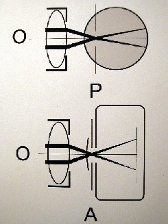
Figure 1. (top) The aperture of the observer eye, the pupil P fits the external aperture of the microscope ocular A. (below) The digital camera's aperture A is coincident with the microscope's external ocular aperture A.
Slit lamp microscope photography, as offered by slit lamp microscope manufacturers, relies on the exchange of one microscope ocular for a special optical camera adapter. It is possible, however, to keep the ocular for photography without a special optical adapter, as has been reported,1,2 providing the camera's optical dimensions are suitable and its optical power is that of the observer eye. Amateur astronomers confirmed this,3 realizing that several types of commercial amateur digital cameras qualify. They are fitted directly to the telescope oculars. The oculars of telescopes use the same optical principle as those of the slit lamp microscope and the fundus camera. This provides sufficient reason to attempt digital photography in ophthalmology for affordable telemedicine.
Method
The placement of the camera's objective lens in line with a microscope ocular succeeds only if the camera optics equals the power of the observer eye (See Figure1). Instead of fitting the the pupil of the observer's eye to the ocular, it is now the "pupil" of the camera that is fitted to the ocular. The required dimension of the camera is its focal length (f:17 mm) that must be within the range of the camera's zoom setting. This provides a camera aperture position for the same plane that the pupil of the observer eye occupies.
 |
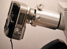 |
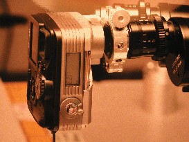
| Figure 2. The mechanical adapter for the digital camera on the ocular of the slit lamp microscope. | Figure 3. The mechanical adapter for the digital camera attached to the ocular of the fundus camera. |
I have used an Olympus Camedia D-40 Zoom camera for this project with an adjustable zoom range of focal lengths from f:7.25 mm to f:20.3 mm, which includes f:17 mm, the power of the eye. Digital cameras of similar optical dimensions are equally suitable. An observer screen is an essential feature of any digital camera to judge the image prior to the take.
The use of 35-mm film photography relies on an electronic flash to shorten the exposure time to a minimum. This is important for photography of the eye, which is essentially a moving target.
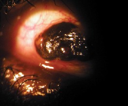 |
| Figure 4. Anterior segment photography: Large conjunctival nevus. |
The method of digital photography without electronic flash that I describe also depends on short exposure by a combination of intense illumination, the inherent high light sensitivity of digital photography, and on an open camera aperture. Prior to the recording, the choice is made from the program settings of the camera. It is useful to set the aperture open so that the high luminosity provides the best condition for the shortest possible exposure time. The recorded average should be 1/100 of a second or less. Several takes are recommended for the best choice of the digital image record.
Although manually holding the digital camera to the ocular is possible with acceptable results, a mechanical device to keep the camera aligned to the ocular is preferred (See Figures 2, 3). This frees the observer to adjust the combined equipment precisely prior to the taking of the picture.
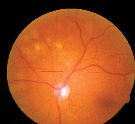 |
| Figure 5. Fundus photography of laser photocoagulation for diabetic retinopathy through the ocular of the fundus camera. |
For the posterior segment, the Gullstrand system of the fundus camera is most suitable. Any fundus camera observer ocular is suitable for this type of digital photography (See Figure 3). This upgrades a fundus camera designed for photography on film to become an affordable digital image camera (See Figure 5, 7). Images are stored on computer memory to transmit over the Internet.
Stereo-paired images (See Figure 6) are juxtapositioned on the computer screen with the help of suitable programs (Microsoft Word, Picture It, Photoshop). This provides the viewer with a split-screen
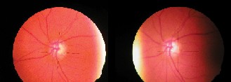 |
| Figure 6. 3D optic nerve cupping with stereoimaging on computer. For spontaneous stereo viewing with overconvergence or prism device (8 D OU prism base in). |
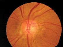 |
| Figure 7. Disc details in small field with large magnification. |
Digital photography with direct recording through the microscope's observer oculars offers a cost-effective method of digital imaging of the anterior and posterior segment of the eye. It offers an upgrade of existing equipment for the purpose of telemedicine through any office computer that connects with the Internet.
Dr. Schirmer is the chairman (emer.) of ophthalmology at St. Mary's Hospital, Department of Ophthalmology, and an assistant professor at McGill University. He holds several patents on afocal microscopy and laser attachment as well as on laser delivery through diagnostic contact lenses, but has no commercial interest in the devices or in the method described. He gratefully acknowledges C. Bryson's help with computer imaging.
1. Schirmer KE. Microfilm Photography in Ophthalmology. Brit.J.Ophthalmol. 1965;49:76-79.
2. Schirmer KE, Jacques LY. Microscope ocular fundus photography. Ophthalmologica (Basel).1987; 195:81-83.
3. Astronomy Products, Scopetronics; scopetronics.com


