Presentation
A 75-year-old Caucasian male presented with one week of painful, progressive, constant right blepharoptosis associated with a right-sided headache. The patient denied changes in vision, diplopia, fever, jaw claudication, scalp tenderness or proximal muscle weakness.
Medical History
Past ocular history was unremarkable. Past medical history included atrial fibrillation, type II diabetes mellitus and hypertension. He also previously had prostate adenocarcinoma managed by radical prostatectomy in 2005, followed by radiotherapy for recurrence in 2006 and enzalutamide (anti-androgen) therapy. In 2012, the patient developed metastases to the spine for which he received palliative external beam radiotherapy. Family history and social history were non-contributory.
Current medications included metoprolol, digoxin, glimepiride, atorvastatin, omeprazole, leuprolide and enzalutamide.
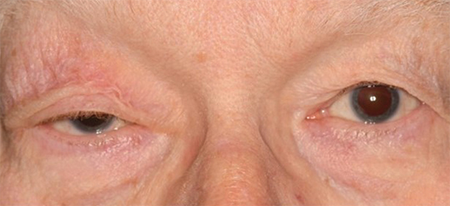 |
| Figure 1. External photograph showing blepharoptosis of the upper eyelid. |
Examination
On examination, best corrected visual acuity was 20/40 OD and 20/25 OS. Pupils were normal, and confrontation visual fields were full in both eyes. Extraocular motility demonstrated a 20-percent limitation of upgaze of the right eye, but was otherwise normal. External examination was remarkable for 3 mm of non-fatigable right upper eyelid ptosis (Figure 1). There was no proptosis by Hertel exophthalmometry and no hypesthesia. Color plates were 7/8 OD and 8/8 OS.
Anterior segment examination showed an age-related nuclear sclerotic cataract OU, but was otherwise unremarkable. Dilated fundus examination demonstrated a blonde fundus with macular drusen in each eye. The cup-to-disc ratio was 0.4 OU without disc edema or pallor.
What is your diagnosis? What further workup would you pursue?
Please click this link for diagnosis, workup, treatment and discussion.


