WITH PHAKIC INTRAOCULAR LENSES becoming increasingly popular, the importance of being able to quickly and accurately measure the anterior chamber has grown. Now, a new optical coherence tomography anterior segment scanner from Carl Zeiss Meditec (Dublin, Calif.), currently in clinical trials, shows promise as an easy and accurate way to do just that.
The new instrument is called the Visante OCT. (Visante is an amalgam of "vision" and "anterior.") It uses 1310-nm infrared light to visualize a "slice" through the anterior chamber. Unlike the 820-nm light used by the Stratus OCT, the longer wavelength readily scans through the sclera and iris, making it particularly useful for the assessment of narrow angles and measuring anterior chamber width.
Advantages and Limitations
The Visante OCT is designed to image the shape, size and position of anterior components and make precise measurements of the distances between them, including angle-to-angle, angle size in degrees, pupil diameter, anterior chamber depth, and thickness and radii of curvature of the crystalline lens. The Visante"s software eliminates any measurement distortion induced by optical transmission factors. It captures the entire anterior segment in one step, with a resolution of about 10 µm.
|
|
|
GEORGES BAIKOFF, MD |
|
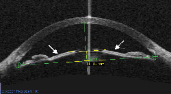
In this Visante OCT scan, iris tissue (marked by arrows) is compressed between an implanted Artisan lens and the patient"s crystalline lens, resulting in pigment dispersion. Note the "crystalline lens rise" of 849 µm ( a useful measurement defined by Georges Baikoff, MD). |
The instrument"s primary limitation is that pigmentation on the posterior side of the iris blocks the penetration of infrared light. That means that in most eyes, the Visante can"t measure sulcus to sulcus.
Noteworthy features include:
- The Visante OCT is non-contact. This minimizes patient discomfort, prevents chamber distortion, reduces the time required to perform a scan, and eliminates cleaning and disinfecting.
Use With Phakic IOLs
In addition to providing valuable preop and postop information about the anterior segment, the Visante OCT may help address several specific concerns:
In addition to providing valuable preop and postop information about the anterior segment, the Visante OCT may help address several specific concerns:
- Iris pigment dispersion. Implantation of the Verisyse phakic lens, which is clipped to the front of the iris, has triggered iris pigment dispersion in some eyes. Work by Georges Baikoff, MD, using the Visante OCT, has shown that this pigment dispersion is a function of how much the iris tends to bow forward toward the cornea, rather than the anterior chamber depth.1 In some patients, scans have found the iris compressed between the crystalline lens and the phakic IOL (see page 33). The Visante can help weed out patients whose irises are likely to be a problem.
|
|