In this column, my goal has always been to speak to my friends and colleagues about the incredible gift and opportunity we have as ophthalmologists to recognize disease and its underlying causes and sometimes be able to use this understanding to intervene therapeutically. After all these years I am still in awe of our collective ability to treat disease with our current knowledge, while exploring the unknown for better interventions. As a clinician, I strive to combine the critical observation skills described by Sir William Osler as, “Listen carefully to the patient, he is trying to tell you his diagnosis,” and the integrative effort highlighted by Arthur Conan Doyle who, as it turns out, was also a physician. The deductive reasoning exemplified by the fictional Sherlock Holmes has guided me in looking for the clinical pearls I tried to imbed in these columns. With this, my final column, I’ll share some parting tips.
Looking Back
The expanding scope of our knowledge, of biochemical pathways, cellular biology and genetic modulation, has continued to accelerate over the years, and I must admit I have used the monthly deadlines and selected topics to keep myself, as well my readers, abreast. I will continue to read a wide range of scientific literature and hope you will too. But it is at these times I start to reel off all that is still left unsaid ...
Please consider debriding corneal ulcers, the removal of fibrin and bacterial load and necrotic tissue will enhance antibiotic kinetics. Debridement and ubi pus, ibi evacuo have stood the test of time, back to the Romans. Then a drop of peroxide; don’t forget the value of disinfectants in this era of uncertain antibiotic sensitivity. I’ve also noted that frequent use of topical aminoglycosides such as gentamicin dramatically decreases melt in scleromalacia. I have no idea why. I was always waiting for a larger series to report this.
When I approached him about it, corneal expert Claes Dohlman, MD, PhD, agreed that the
 |
My use of topical leukotriene beta 4 produced a prolonged migraine and a massive white blood cell infiltration into conjunctiva—fortunately without degranulation stimulus. Even this adverse reaction provided a kernel of wisdom: Isn’t it reasonable to suggest that a migraine associated with throbbing pain over one eye without visual disturbance, identified and slightly relieved by pressure on a pulsating temporal artery, might be related to pollen entering the eye in a predisposed (allergic) subject? Under this premise, allergic neutrophil infiltration results in local degranulation, release of histamine and other vasoactive amines, and pathologic vasoconstriction. Histamine had already been identified in Horton’s cluster headaches. So, in hindsight, after putting the most powerful neutrophil chemotactic factor, LTB4, in my right eye, it was no wonder I developed a severe migraine for more than a week. At that point, I thought that I had done myself in! The use of beta-blocker drops and topical high-strength ketotifen eye drops provided significant relief and became staples among our type-one personality, migraine-sensitive scientists.
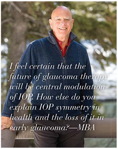
At this time we still don’t fully understand the cause of open-angle glaucoma and the full mechanism of treatments like alpha-agonists. Parasympathetic agents, beta blockers and prostanoid therapies point to a large number of possible etiologies. But I feel certain that the future will be central modulation of IOP. How else do you explain IOP symmetry in health and the loss of it in early glaucoma? All other paired organs in the body are under central control. When I put either a beta blocker or an alpha agonist in my nose (just one nostril), the IOP drops in both eyes: with this N of 1, I can rest my case. Those of us working on arachidonic acid knew prostaglandins could reduce pressure in inflammation; we called it hypotony and thought it would lead to phthisis bulbi. Even though I put these agents in my eye, it took a non-ophthalmologist to see its potential use in glaucoma.
I cannot help wondering what we might learn by looking at other ultrafiltration-producing organs, the semicircular canal fluid, cerebral spinal fluid and perhaps aspects of the glomerulus. Secretion of an acellular, protein-free fluid is not unique to the aqueous humor. Certainly the ciliary body and choroid plexus share some physiology and pathology.
Team Efforts
I have learned much with Paul Gomes (Vice President for Allergy at the ophthalmology consulting firm Ora Inc.) throughout decades of challenging tens of thousands of eyes with pollen. Twenty FDA drug approvals aside, many questions remain unanswered, particularly as they relate to chronic or panseasonal ocular allergy. Representing perhaps one-third of all ocular allergy patients, they don’t respond well, if at all, to current antihistamine or mast-cell stabilization therapy. There’s a need to understand how these patients differ immunologically from others with histamine-dependent allergy. Of course, ongoing research will point the way and clinical trials will continue. I have no doubt there will be another generation of anti-allergy eye drops.
Vernal keratoconjunctivitis has been of great interest to me. The dramatic effect of aspirin, particularly in contrast to the negligible effect of other NSAIDs in this disease, suggests additional ocular anti-inflammatory effects of this ancient, tree-bark-derived agent. In a human arachidonic acid ocular challenge model it proved to be far superior to NSAIDs. Asprin is difficult to formulate for topical use, but new nanotechnology formulations may change that. Another key aspect of VKC is that it proved to be a great model for understanding the role of histamine in the eye; the absence of histaminase appears to be a primary underlying etiology of VKC.1 This implies a metabolic disease with delayed development of this enzyme (as VKC patients invariably outgrow the disease) allowing an allergic reaction to run wild. The path for therapeutic development seems clear and I hope someone takes this route.
More Connections
Dry eye is undoubtedly a wide range of conditions and diseases still grouped together and, in my opinion, not driven by subtle changes in osmolality. New therapies in the past two years show a role for treating inflammation and neural lethargy, and we at Ora have been pleased to be part of these and other, up-and-coming dry-eye treatments. Meibomian gland “disease” must be separated from dramatic gland dropout in normal aging and the life cycle of individual holocrine glands that puts them through an inspiration phase. We have enough diseases without creating more.
We have not made dramatic progress on anti-inflammatory therapy since the advent of steroids in the eye in the mid-1950s. “Soft steroids” was marketing hype for “weak steroids,” and have still not broken the anti-inflammation and intraocular pressure effect connection. Cyclooxygenase-1 inhibitors have helped in the margins. Topical aspirin could hold some benefit if formulation difficulties can be addressed, and the splenic tyrosine kinase inhibitors that target the immunoglobin-receptor pathways hold great promise for the future.2
As for wet age-related macular degeneration, I once told my colleague Judah Folkman, MD, “Anti-VEGF will not work and will only produce necrosis.” I was wrong. Had he lived, he would have been a Nobel Laureate.
The next great frontier is dry AMD, which is currently the center of much attention. The recognition that form follows function is leading to exciting preliminary results in psychophysical tests for early identification of dry AMD at the reversible stage.3 I confess to being focused on this disease with my own research group at Ora, as we aim to develop new models for screening.
Although the chimeric antigen receptor therapy revolution is upon us, changes in drug reimbursement do not bode well for the continued investment in research needed for the next generation.4,5 In ophthalmology, there is no longer interest in incremental drug development, which has been at the core of our historic progress, thanks to the economic benefits accruing to the six sisters, the large pharmacy chains and their benefit managers. These nefarious managers control drug availabilities even after Food and Drug Administration approval of new agents with demonstrated superiority to existing agents, in order to preserve a multi-billion dollar rebate scam that resembles the payola debacle in the music industry 50 years ago. In time, this will get the legislative attention it deserves and then, hopefully, the drug development that has increased the length and quality of our lives will continue apace.
The routine eye exam is now quite clearly capable of being fully automated, with any initial abnormality tagged for referral. This will most certainly shift ophthalmic practice to a pathology-enriched environment and enhance early detection of disease and visual dysfunction; the compiling, organizing, sequencing and miniaturizing of appropriate equipment are well under way. Computerized recognition of retinal and corneal abnormalities is now excellent and having had the chance to help in their clinical regulatory progression, I find them impressive. I suspect these kiosks will be placed in pharmacies and dispense prescriptions for glasses and contacts with minimal or no charge for testing.
Artificial intelligence is much in the news today and its application to clinical history is burgeoning. In an early version smart form, 25 years ago, we developed a questionnaire/algorithm for differentiating peripheral immune infiltrates from early infectious corneal ulcers, which proved important in the early years of continuous-wear contact lenses. This form then proved in thousands of cases to be more accurate than any one of us over time. The prolonged nature of that endeavor for one situation can be obviated by self-learning questionnaires and their associated algorithms, leading ultimately to timely diagnosis online from a mobile device. The Kardia smartphone application (AliveCor, Mountain View, Calif.) allows for a remote electrocardiogram completed through a small touch pad connected via Bluetooth to a user’s smartphone, then to a computer for analysis. Abnormalities are sent to a live cardiologist, for further examination. At Ora, we have already validated and used (in FDA trials) a smartphone camera with an adapted eye cone to control quality, and an alarm-prompted, smartphone-based treatment diary. Embrace the future!
Finally, my thanks to many colleagues, particularly Mathea Allansmith, Wiley Chambers, Claes Dohlman, Joan Miller, Jerry Cagle, Art Neufeld and the late Sean Murphy and David Maurice. Your guidance, support and friendship over half a century have been much appreciated. I conclude as I have in many lectures, in many countries, over many years, with one of my favorite pearls: If it itches, it’s allergy; if it burns, it’s dry eye; and if it sticks in the morning, it’s bacterial. REVIEW
Dr. Abelson is a clinical professor of ophthalmology at Harvard Medical School and Emeritus Surgeon and Trustee at the Massachusetts Eye and Ear Infirmary. He is the founder of the ophthalmic consulting firm, Ora.
1. Abelson MB, Leonardi AA, et al. Histaminase activity in patients with vernal keratoconjunctivitis. Ophthalmology 1995;102:12:1958-63.
2. Geahlen RL. Getting Syk: Spleen tyrosine kinase as a therapeutic target. Trends Pharmacol Sci 2014;35:8:414-22.
3. Owsley C, Huisingh C, Clark ME, et al. Comparison of visual function in older eyes in the earliest stages of AMD to those in normal macular health. Curr Eye Res 2016;41:2:266-72.
4. Hartmann J, Schüßler-Lenz M, Bondanza A, Buchholz CJ. Clinical development of CAR T cells-challenges and opportunities in translating innovative treatment concepts. EMBO Mol Med 2017 Aug 1. pii: e201607485.
5.https://www.novartis.com/news/media-releases/novartis-receives-first-ever-fda-approval-car-t-cell-therapy-kymriahtm-ctl019. Accessed 30 Aug 2017.


