Two hundred columns fly by before you know it. We started writing this monthly department back in 1994 with the aim of helping to keep clinicians up-to-date on the latest in therapeutics and therapeutic thinking. It was about that time that a new trend in medical "philosophy" was gaining momentum: evidence-based medicine. The scientific method was well established in basic science laboratories, but it wasn't until 1993 that the first evidence-based medical practice guidelines were published.1 After those guidelines were published, the proverbial floodgates opened, ushering in many changes in medical training (the transformation from "discipline-based" to "case-based") and putting an increased emphasis on the clinical trial as a source for treatment rationale. In this setting, the idea of translational medicine—where the emphasis is on bringing basic science findings to clinical practice—became a central tenet in ocular drug development. This month's column will describe some examples of how translational medicine has changed ophthalmic drugs over the past 16 years.
In the years before we started writing Therapeutic Topics, the available therapies for treating ocular allergy were hit-or-miss at best. We did studies that helped to define mediators of allergic inflammation2 and the role of histamine receptor subtypes,3 and in the course of these and other studies we realized that the process of developing new treatments was hit-or-miss, as well. This led to the development (or, more accurately, refinement) of the conjunctival allergen challenge model for clinical assessment of ocular allergy therapies.4,5
The CAC was a significant advance for several reasons. It provided a way to control allergen concentration, allergen exposure and drug treatments simultaneously. This control allowed us to titrate pollen exposure to evoke predictable levels of both signs and symptoms of the conjunctival response. In addition, we developed a standardized grading system employed by a trained clinician. These aspects of the model provide the means to elicit an allergic response that is robust and reproducible. In this setting, evaluation of new therapeutics became an objective, quantitative procedure. Many drugs, including olopatadine, levocabastine and alcaftadine, have been tested and proven effective using this paradigm. The overall result is a significant improvement in the standard of clinical care for ocular allergy.
 At times the pace of scientific progress is slowed by conventional wisdom; in these instances it's sometimes necessary to reconsider generally accepted methods and assumptions.
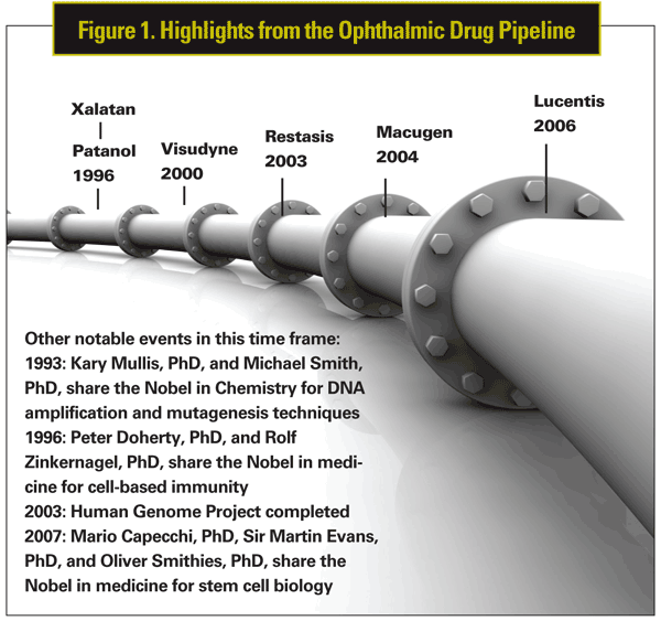
For instance, clinicians are familiar with instances of hypotony that can accompany ocular inflammation. Few, if any ever consider what connection there may be between these two conditions. Two who did were Laszlo Bito, PhD, and Carl Camras, MD, the original creators of the paradigm-shifting glaucoma drug Xalatan (latanoprost, Pfizer). Drs. Bito and Camras thought that the hypotony might be related to some autocoid (such as a prostaglandin) produced during an inflammatory response. In a 1977 publication they described the results of elegant experiments in rabbits that suggested prostaglandins do not work by "alterations in secretory mechanisms or to pseudofacility."6 Their data suggested that prostaglandin-mediated reductions in intraocular pressure could be "attributed to a reduction in true outflow resistance" within the trabecular network.6 This led them to hypothesize that some prostaglandins and their analogues could be useful in the treatment of ocular hypertension or glaucoma.6
Lucky for us, Drs. Bito and Camras were correct. They continued their research with latanoprost, a prostaglandin F2-alpha analogue, into the next two decades7,8 and in 1996 Xalatan was approved by the Food and Drug Administration. Since then, prostaglandin analogues have demonstrated improved efficacy and superior safety profiles compared to older glaucoma medications such as carbonic anhydrase inhibitors, alpha-adrenergic agonists and beta-blockers. They are now considered first-line therapies for open-angle glaucoma and, as hypothesized in 1977, are believed to work by increasing uveoscleral fluid outflow and reducing intraocular pressure on the optic nerve.
The impact of the translational approach in ophthalmology is also exemplified by the development of pegaptanib sodium (Macugen; Eyetech Pharmaceuticals/Pfizer)—a modified RNA aptamer molecule indicated for treatment of neovascular age-related macular degeneration.9 Among the distinguishing attributes of pegaptanib sodium is that it became the first aptamer successfully developed as a clinical therapeutic agent when it was approved for the treatment of AMD in 2004.
The story of pegaptanib began in the 1990s when Drs. Anthony Adamis and David Guyer began their research into retinal angiogenesis as a cause for disease. At this time, basic research had shown that ocular neovascularization is regulated by diffusible growth factors including vascular endothelial growth factor. Studies found that under hypoxic conditions (which are conditions analogous to vascular occlusion and AMD), VEGF levels became elevated in retinas.10 Based upon this discovery, Drs. Adamis and Guyer recognized that a blockade of VEGF and factors promoting neovascularization were likely therapeutic targets.
In 2000 Eyetech was founded with the goal of characterizing and developing a new chemical entity: an RNA aptamer named NX1838. At Eyetech, Drs. Adamis and Guyer were largely responsible for pursuing the molecule (later renamed pegaptanib) as a therapeutic agent by determining its preclinical activities11 and working towards its successful clinical development.12 It's important to note, however, that pegaptanib wouldn't have been possible without Craig Tuerk, PhD, and Larry Gold, PhD, who developed a technique known as systematic evolution of ligands by exponential enrichment or SELEX.13 Their SELEX technique leverages naturally occurring, highly specific binding interactions between oligonucleotides and their cellular or extracellular targets.13 Dr. Gold and coworkers at NeXstart Pharmaceuticals used SELEX to identify and develop the NX1838 aptamer (before it was purchased by Eyetech), which participates in highly specific binding interactions with the VEGF-165 isoform. Preclinical characterization of NX1838 demonstrated that it was capable of blocking VEGF-165 extracellularly and abrogating VEGF-induced neovascularization and vascular permeability.14
The development of pegaptanib was critical in validating the VEGF pathway as a clinical therapeutic target for AMD and other retinal vascular diseases. In December 2004, when pegaptanib was approved by the FDA for the treatment of wet AMD, patients had few therapeutic options: Laser photocoagulation therapy or Visudyne (verteporfin for injection, QLT Ophthalmics) photodynamic therapy were used to reduce choroidal neovascularization in advanced exudative AMD patients.15 Since then, other drugs targeting VEGF have proven vital as AMD therapies, including the anti-VEGF monoclonal antibody fragment Lucentis (ranibizumab, Genentech).
But what research is paving the way for new therapeutics to treat conditions affecting the front of the eye? In the fields of allergy and dry eye, a focus of basic science in recent years has been on the humoral and cellular control of inflammation. The importance of the immune system in ocular allergy is nothing new, but there's a growing consensus that the many disparate pathologies of dry-eye disease are united by immune-system based etiolologies.16
A 1998 review article by Baylor College of Medicine's Stephen Pflugfelder, MD, was one of the first publications to describe a role for inflammation in dry-eye disease.17 Several laboratories had found evidence of T-cell involvement in types of dry eye, and Dr. Pflugfelder's research group published a series of studies showing increased cytokines, chemokines and inflammatory markers within the tears of dry-eye patients.18-20 As a follow-up to this, they showed that inflammatory markers can be induced in animal models of dry eye.21 At about this time, Renee Kawsan, DVM, and her colleagues, who had conducted studies on keratoconjunctivitis sicca in dogs,22 published the results of a trial using topical cyclosporine, a T-cell immunosuppressant used in organ transplants. In their study cyclosporine had two effects: it reduced granulation and corneal scarring, and it increased tear production. Within three years cyclosporine (Restasis, Allergan) was approved by the FDA as the first therapeutic agent for the treatment of dry eye.23 With this drug approval came an expectation that more therapies would soon follow—so what happened?
A number of factors have slowed the progression of dry-eye therapeutics from researchers' work at the lab bench to the clinic. One factor is that while the success of cyclosporine was indicative of immune-based mechanism for dry-eye disease, the drug is only effective in about 15 percent of all patients. In addition, the ability of cyclosporine to act as an immune-modulator is inconsistent with the slow onset observed for the therapeutic effect on dry eye. This could reflect some other action of cyclosporine, the chronic nature of the disease or some combination of factors.
Collectively, these issues prevented cyclosporine from becoming a "first-in-a-class" drug for dry eye. No other drugs have been approved for use in dry eye since the Restasis approval in 2003. And while the immunosuppressant glucocorticoids have reliably proved effective in dry-eye disease, their use is limited by the adverse effects associated with chronic steroid therapy (currently, no steroids are FDA-approved for dry eye).
Another factor that has slowed development of new therapeutics for dry eye is the difficulty in establishing robust clinical endpoints to confirm or refute the efficacy of test compounds. Many early dry-eye studies were based solely on subjective measures of symptoms, and the difficulties associated with such measures are well documented.24 The bar is set higher for dry-eye disease than most other disorders because the only valid placebo comparator (artificial tears) is itself a recommended treatment for dry eye. So while the unmet need in dry eye is great, so too is the difficulty in establishing a record of solid clinical efficacy for drugs that, in theory, appear to be good candidates for dry-eye therapy.
This takes us back to the CAC model for allergic conjunctivitis. What we have learned from CAC clinical studies can be applied to dry-eye disease models, as well.25 Our goal is to improve existing diagnostic tools and to refine and expand our clinical endpoint measures. For example, we have developed a controlled adverse environment model for clinical assessment of dry-eye therapies. In this model, subjects sit in a closed chamber and we manipulate environmental conditions (temperature, humidity and air movement) to elicit signs and symptoms of dry eye.
We have also developed new methods to quantify the clinical signs of dry eye. One measure, the ocular protection index, is a ratio calculated from tear-film breakup time and the inter-blink interval. OPI provides a measurement of corneal exposure and a more objective assessment of the role of tear-film stability in dry-eye disease. A second measure is inter-blink interval visual acuity decay, a computerized assessment of visual acuity dynamics in the CAE setting. The IVAD is useful because it provides an objective way to measure blurred vision and loss of visual acuity in subjects with dry eye.
We know it's critical to apply the same scientific rigor that we use in preclinical studies when we design and validate a clinical model. We also know that the selection and screening of subjects is critical; if study subjects don't provide an accurate representation of the clinical features of the disease then they can't be expected be useful in the assessment of potential therapies. In short, we have to keep in mind that the practice of translational medicine is an endeavor that succeeds when the scientific method is applied throughout the process.
Dr. Abelson, an associate clinical professor of ophthalmology at
1. Sox HC, Woolf SH. Evidence-based practice guidelines from the
2. Udell IJ,
3. Abelson MB, Udell IJ. H2-receptors in the human ocular surface. Arch Ophthalmol 1981;99:2:302-4.
4. Abelson MB, Chambers WA, Smith LM. Conjunctival allergen challenge. A clinical
approach to studying allergic conjunctivitis. Arch Ophthalmol 1990;108:1:84-8.
5. Abelson MB. Comparison of the conjunctival allergen challenge model with the environmental model of allergic conjunctivitis. Acta Ophthalmol Scand Suppl 1999;228:38-42
6. Camras CB, Bito LZ, Eakins KE. Reduction of intraocular pressure by prostaglandins applied topically to the eyes of conscious rabbits. Investig Ophthamol
7. Camras CB Schumer RA, Marsk A, et al. Intraocular pressure reduction with PhXA34, a new prostaglandin analogue, in patients with ocular hypertension. Arch Ophthalmol 1992;110:1733-1738.
8. Camras CB,
9. Ng EW, Shima DT, Calias P, et al. Pegaptanib, a targeted anti-VEGF aptamer for ocular vascular disease. Nature Rev 2006;5:123-32.
10. Miller JW, Adamis AP, Shima DT, et al. Vascular endothelial growth factor/vascular
permeability factor is temporally and spatially correlated with ocular angiogenesis in a primate model. Am J Pathol 1994;145:3:574-84.
11. Carrasquillo KG, Ricker JA, Rigas IK, et al. Controlled delivery of the anti-VEGF aptamer EYE001 with poly(lactic-co-glycolic) acid microspheres. Invest Ophthalmol Vis Sci 2003;44:290–9.
12. Macugen AMD Study Group. Pegaptanib 1-year systemic safety results from a safety–pharmacokinetic trial in patients with neovascular age-related macular degeneration. Ophthalmology 2007;114:1702–12.
13. Tuerk C, Gold L. Systematic evolution of ligands by exponential enrichment: RNA ligands to bacteriophage t4 DNA polymerase. Science 1990;249:4968:505-10.
14. Ruckman J, Green LS, Beeson J, et al. 2'-Fluoropyrimidine RNA-based aptamers to the 165-amino acid form of vascular endothelial growth factor (VEGF165). J Biol Chem1998;273:32:20556–20567.
15. Schmidt-Erfurth U, Miller JW, Sickenberg M,et al. Photodynamic therapy with verteporfin for choroidal neovascularization caused by age-related macular degeneration: Results of retreatments in a phase 1 and 2 study. Arch Ophthalmol 1999;117:9:1177-87.
16. Stern ME, Schaumburg CS, Dana R, et al. Autoimmunity at the ocular surface: Pathogenesis and regulation. Mucosal Immunol 2010;3:425-442.
17. Pflugfelder, SC. Anti-inflammatory therapy for dry eye. Am J Ophthalmol 1998;137:337–342.
18. Pflugfelder SC, Jones D, Ji ZH, et al. Altered cytokine balance in the tear fluid and conjunctiva of patients with Sjogren's syndrome keratoconjunctivitis sicca. Curr Eye Res 1999;19:201-211.
19. Solomon A, Dursun D, Liu ZG, et al. Pro- and anti-inflammatory forms of interleukin-1 in the tear fluid and conjunctiva of patients with dry-eye disease. Inv Ophth
20. Pflugfelder SC, Tseng SCG, Sanabria O, et al. Evaluation of subjective assessments and objective diagnostic tests for diagnosing tear-film disorders known to cause ocular irritation. Cornea 1998;17:38-56
21. Luo LH, Li DQ, Doshi A, et al. Experimental dry eye stimulates production of inflammatory cytokines and MMP-9 and activates MAPK signaling pathways on the ocular surface. Inv Ophth
22. Kaswan RL,
23.Restasis (cyclosporine ophthalmic emulsion) 0.05% Prescribing Information. Allergan, Inc.,
24. Smith J, Nichols KK, Baldwin EK. Current patterns in the use of diagnostic tests in dry-eye evaluation. Cornea 2008;27:656-62.
25. Ousler GW, Gomes PJ, Welch D,


