Three factors are fueling what some specialists are calling a potential retina epidemic: the increased use of anti-VEGF treatments, the aging population and the increased incidence of obesity. With all three expected to rise for the foreseeable future, the result is that retina specialists need to see more patients more often.
The Makings of an Epidemic
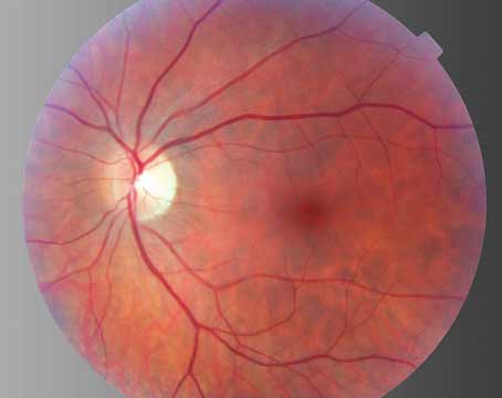
Anti-VEGF therapy is used for several retinal conditions, and many patients require frequent injections. “One of the major prevalent diseases for retina-related evaluations has been age-related macular degeneration,” says Timothy G. Murray, MD, from the Bascom Palmer Eye Institute in Miami. “Traditionally, we’ve had very few treatment options available for AMD, particularly for patients with wet AMD. The introduction of anti-VEGF treatments has pivotally changed our practices, because this whole group of patients who existed but had no care options now have moved into a treatable disease profile.”
Before the introduction of anti-VEGF therapy, these patients may have only been seen in the office once a year. Now, they may be seen as often as monthly. “So, your practice volume in terms of the clinical impact increased 12-fold,” Dr. Murray says. That heralds what we are going to potentially see going forward with other diseases, such as proliferative and nonproliferative diabetic retinopathy. For these conditions, we often would treat once, and then we would assess response to treatment in two to four months. In many practices, that response to treatment was assessed in four months. When those diseases became amenable to anti-VEGF therapy, where the clinical trials have focused on a one-month interval to treatment, patients whom you may have been seeing twice a year or three times a year are now seen 12 times a year. As you rule conditions into treatment regimens that require active intervention at short intervals, even without changing the incidence or prevalence of the disease, you greatly change its impact on the retina practice.”
John Thompson, MD, a retina specialist practicing in Towson, Maryland, agrees. “Retina specialists have become very busy during the past six years with the availability of the anti-VEGF drugs Lucentis and Avastin. We thought we could give these treatments monthly for a while, and then less often, and then stop, but it has become increasingly apparent that these treatments for macular degeneration have to be given in most patients for a long time, often monthly or every other month.”
Central retinal vein occlusion and branch retinal vein occlusion can also be effectively treated with anti-VEGF therapy. “All of these conditions are incredibly prevalent,” Dr. Murray says.
Many of these conditions are age-related, so the epidemic is just beginning. As the baby boom generation becomes older and life expectancy is extended, more patients will require treatment.
Additionally, the population is changing its environmental and life exposures. “As people become progressively more obese, which we have seen over the past several decades, we become at risk for diseases associated with that, which includes obesity-related hypertension, diabetes, and cardiovascular disease, all of which have a huge impact on a retinal practice,” Dr. Murray explains.
Preparing Your Practice
To accommodate seeing more patients more often, retina specialists will need to become more efficient and possibly add more space to their practices. “Doctors will need to streamline their operations to try to make themselves more efficient and to use technicians to do some of the other work that doesn’t require a physician,” Dr. Thompson says. “The initial response of most retinal specialists has been to use paraprofessionals in our offices to help us to see patients more efficiently. I am not aware of any general ophthalmologists doing anti-VEGF injections. However, this may be an option for patients in areas without a retinal specialist so they wouldn’t have to travel a long distance. There hasn’t been a lot of interest on the part of general ophthalmologists for doing these injections.”
Some retina specialists have had to enlarge or reconfigure their office facilities. “They have had to increase the number of examination rooms to allow them to handle more patients, and many practices have had to hire additional staff to accommodate the patient flow,” Dr. Thompson adds.
|
In Dr. Murray’s practice, a certified ophthalmic technician does the prescreening. This technician tests vision, refraction and intraocular pressure and takes the initial patient history. The technician also dilates the eye. Dr. Murray notes that his practice provides ancillary testing. “At most retina practices now, that includes photographic and OCT documentation of the retina, which is performed by a technician,” he says. “Additional testing like ultrasonography is performed by another technician at most large practices. This allows retina specialists to be able to evaluate patients thoroughly and rapidly.” For added efficiency, his practice has standardized documentation for imaging.
The practice has also had to add space to accommodate the increasing patient load. “This can be quite expensive, because it requires that you have more lanes to practice in than you’ve had previously,” he explains. “In the past, some smaller offices may not have had a technician, and the physician has done the entire exam. That was a wonderful interaction with your patient but incredibly inefficient for the physician. In my office, I have three technicians who have their own office lanes that are fully equipped for complete patient evaluation. The flow goes from the technician to the photographer for imaging, to the echographer and then comes back into a separate room for me.”
He continuously goes from room to room seeing patients. “You have to become very good at rapidly assimilating the complexity of data now available to you, and you have to be very good about establishing good rapport with your patients very quickly,” says Dr. Murray. “You need to be able to make a quick diagnosis and institute a treatment plan. We are seeing 70 patients in a day. Because we do so many intravitreal injections, 35 to 40 in a clinic day, I have two injection rooms that are separate and staffed by two nurses. So, we have three rooms for techs, three rooms for the doctor, two rooms for injections, two rooms for photography and two rooms for ultrasound. My single-physician practice has now become huge, and the costs of providing this space, these services, personnel and the equipment are high.”
Unfortunately, the revenues for injections and imaging are not high. Dr. Murray and his colleagues recently published an evaluation of economic efficiencies in clinical retina practice.1 In this study, activity-based costing analyses were performed at two different types of retinal practices in the United States. One practice was a small, single-specialty group practice, and the other was Bascom Palmer Eye Institute, an academic, hospital-based practice. The study found that the small practice outperformed the larger practice on almost all markers of efficiency. At Bascom Palmer, only four service lines were profitable: nonlaser surgery, laser surgery, non-OCT diagnostics and injections. Profit margins ranged from 62 percent for nonlaser surgery to 1 percent for intravitreal injections. Office visits and OCT imaging were associated with the largest negative profit margins.
The financial burden can be huge. “The cost of Lucentis for a year, given every four to six weeks, is substantial and runs in the tens of thousands of dollars,” says Martin Friedlander, MD, PhD, chief of the Retina Service in the Division of Ophthalmology at the Scripps Clinic in La Jolla, Calif. “As the population ages and the number of individuals with AMD increases, that number will increase by millions. We need to find alternative ways of delivering the drugs that will decrease the frequency of dosing or we need to find more cost-effective treatments.”
The Future
Alternative treatments for these conditions are currently under investigation. “Combination therapies (which are more effective and last longer), sustained-release drug delivery devices (like nanoparticles or encapsulation cell-based therapies), or treatments that target novel angiogenic/vascular permeability pathways would go a long way in helping out,” Dr. Friedlander notes.
|
Dr. Friedlander believes that future treatment will consist of combination therapy. “I’m talking about targeting multiple angiogenic pathways that play a role in facilitating abnormal blood vessel proliferation of the type we see in macular degeneration,” he says. “Combination therapies, cell-based therapies, and maybe cell-based therapies in which the cells are actually programmed to deliver molecules that would enhance the anti-antigenic effect or that would provide trophic support/rescue for neurons and vasculature may be the answer.”
For example, a recent study conducted at Bascom Palmer evaluated the efficacy of different dosing paradigms of PF-04523655 (an RNAi therapeutic candidate that has been in two Phase II trials for diabetic macular edema and AMD) compared with ranibizumab in patients with neovascular AMD.2 In this study, 151 patients were randomized to one of five treatment groups. All groups received ranibizumab 0.5 mg at baseline. Then, one group received 1 mg of PF every four weeks from week four to week 12; one group received 3 mg of PF every four weeks from week four to week 12; one group received 3 mg of PF every two weeks from week four to week 12; one group received 1 mg of PF plus ranibizumab every four weeks from baseline to week 12; and one group received ranibizumab every four weeks to week 12. All treatments were given as intravitreal injections. At week 16, the combination treatment group achieved greater improvement in mean best-corrected visual acuity than the ranibizumab group (9.5 letters compared with 6.8 letters), and no safety concerns were reported.
According to Dr. Murray, the long-term fix is to train more retina specialists in the future. “That is an excellent strategy, but you need to target the number of people in training to the presumed need,” he says. “A Rand Institute study published in 1995 significantly underestimated the need for ophthalmologists. In fact, at a time when we probably should have been training more ophthalmologists in the United States, we elected to train fewer ophthalmologists. By training fewer ophthalmologists, we trained fewer specialists in retina. Our long-term goal is to match the training environment to the health-care needs of the nation. It’s not a quick fix, however,” he adds. REVIEW
1. Murray TG, Tornambe P, Dugel P, Tong KB. Evaluation of economic efficiencies in clinical retina practice: Activity-based cost analysis and modeling to determine impacts of changes in patient management. Clin Ophthalmol 2011;5:913-925.
2. Nguyen QD, Schachar RA, Nduaka CI, et al; MONET Clinical Study Group. Evaluation of the siRNA PF-04523655 versus ranibizumab for the treatment of neovascular age-related macular degeneration (MONET study). Ophthalmology 2012; Jun 8. [E-pub ahead of print].