Complications during cata-ract surgery have become rare, but when they do occur the surgeon has to know how to proceed. A posterior capsule break can be nerve-wracking for a surgeon whose primary experience is working in the anterior chamber.
Unfortunately, in that situation making poor choices can have serious consequences.
Here, three surgeons—including a retinal specialist and an anterior segment surgeon with extensive retinal experience—share some of their strategies for performing a successful anterior vitrectomy when the capsule unexpectedly breaks during cataract surgery. Our surgeons also weigh in on the debate about whether a cataract surgeon should attempt anterior vitrectomy through a paracentesis or a pars plana incision.
Recognizing the Warning Signs
The first challenge is realizing that the capsule has broken. "Essentially, you get a deepening of the posterior chamber and it goes out of focus," explains Uday Devgan, MD, FACS, chief of ophthalmology at
Louis Nichamin, MD, medical director of the Laurel Eye Clinic in

"Howard Fine has suggested a mnemonic device for remembering three cardinal signs of a ruptured capsule—the three Rs: Room, Rebound loss, and Resistance to phaco," notes Dr. Devgan. "Room refers to increased space in the eye with the deepening of the posterior chamber. Rebound loss refers to the inability of the nucleus to rebound back to a central location after manipulation. Resistance to phaco refers to the surgeon's inability to emulsify the cataract because of prolapsed vitreous blocking the phaco tip."
Allen C. Ho, MD, FACS, who practices at MidAtlantic Retina and the Wills Eye Institute, and is professor of ophthalmology at Thomas Jefferson University School of Medicine in
Setting Up for Vitrectomy
Once it's clear that vitrectomy is necessary, proceed carefully and avoid impulsive actions that could make a bad situation worse.
• Don't pull out the phaco probe. A common first impulse is to pull the phaco probe out of the eye. "This is the worst possible thing to do," notes Dr. Devgan. "When the probe is in there, the infusion is inflating the anterior chamber. That means the pressure in the anterior segment is high while the pressure in the vitreous cavity is medium, so the vitreous remains in the vitreous cavity. But if you pull out of the eye, the pressure in the anterior chamber goes to zero, and the pressurized vitreous will move forward. You'll have vitreous everywhere.
"When the capsule breaks, keep the phaco probe in the eye on position one," he continues. "With your other hand, through your sideport, inject a whole tube of dispersive viscoelastic under the lens fragments, right in front of the vitreous breakthrough. This should push the vitreous posteriorly and the lens fragments up into the anterior chamber. It will also act as a barrier between the anterior segment and the vitreous cavity and repressurize the anterior chamber. Now if you pull out of the eye you've created a barrier, and you still have pressure in the anterior chamber so the vitreous won't prolapse forward."
• Don't chase fragments that have fallen into the vitreous. "You can cause retinal tears by pulling on the vitreous in the process of chasing them," notes Dr. Ho. "Instead, leave the phaco handpiece in the eye and switch to irrigation to maintain the anatomy of the chamber."
• Use the right viscoelastic strategy. Dr. Nichamin says that for the viscoelastic to have the proper effect, placement, technique and choice of viscoelastic all matter. "It's necessary to place the cannula underneath the nucleus and then inject very gently and slowly, as the viscoelastic is pushing both lens fragments and vitreous," he explains.
"You don't want to move the vitreous mass.
"It's also crucial to understand the physical characteristics of the viscoelastic being used," he continues. "The first time I ever used Healon 5 was in this setting; unfortunately I didn't understand its properties adequately. I pushed a fairly large, loose nuclear fragment—one that I could easily have corralled—so far back that I was forced to use the PAL [posterior-assisted levitation] technique to retrieve it.
"Today, depending on the circumstances, I may use as many as three vials of different viscoelastics," he notes. "Use the characteristics of each viscoelastic to your advantage—to maintain space, or to quarantine or compartmentalize the eye. This is not a time to try to save a few bucks."
• Suture the phaco wound before proceeding. "Once you stabilize the anterior chamber, close the phaco wound with a suture," says Dr. Ho. "Anterior vitrectomy is a bimanual technique, so you want to separate the infusion from the anterior vitrectomy probe, one on each side. The only way you can do that and maintain a closed system is to stitch the phaco wound closed."
• Consider providing supplemental anesthesia. "When you're performing an anterior vitrectomy the patient will undoubtedly feel more, and it's going to be a longer procedure," says Dr. Devgan. "So I would supplement with both additional intravenous anesthesia and intracameral nonpreserved lidocaine."
Dr. Nichamin notes that sometimes the surgeon may benefit more from additional anesthesia than the patient. "Sometimes the patient is okay but the doctor is sweating bullets," he says. "If the patient's moving and you want a dead-still eye, a little more sedation or a deeper block can be given. What you don't want to do is feel uncomfortable and rushed in that situation."
However, Dr. Nichamin adds a few caveats. "If you plan to administer a block, seal the incision first," he says. "Ensuring an intact globe will avoid problems related to increased pressure from the block. Alternatively, one can give a little sub-Tenon's supplementation, or deliver a little more intracameral lidocaine. The caveat there is that if the capsule's open, the lidocaine can diffuse back to the retina and cause a temporary amaurosis. Basically, the lidocaine puts the retina to sleep."
The Pars Plana Debate
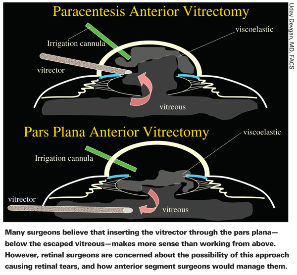
One issue that's become the subject of debate is where the vitrectomy incision should be made when the surgeon is not experienced at working in the posterior segment.
"The pars plana approach makes more sense anatomically; you can pull the vitreous back behind the iris and the capsular rupture," notes Dr. Ho. "My problem with that is that if you go pars plana you need to inspect the peripheral retina and be able to deal with any complications such as a retinal tear or detachment. For that reason, I usually advocate that cataract surgeons who do not have extensive retina training use a limbal approach for an anterior vitrectomy. Many cataract surgeons are not accustomed to inspecting the peripheral retina. And what do you do if you create a retinal tear or detachment?"

Dr. Nichamin has had extensive experience with posterior segment surgery, even though his primary focus today is the anterior segment. "I appreciate the pros and cons on both sides of the argument," he says. "And it's definitely a paradigm shift; working through the pars plana has traditionally been off-limits to the anterior segment surgeon. However, it's become obvious to me that there's no reason anterior segment surgeons should not use a pars plana approach for an anterior segment vitrectomy. It's not particularly difficult, and I think it falls well within the purview of every modern phaco surgeon.
"I don't mean to be cavalier about this," he notes. "It's a new technique that has to be studied and carefully practiced before being used under duress during a live complication. But it offers the surgeon a far more efficient, and I believe much safer, way of performing an anterior vitrectomy.
"It's safer for several reasons," he continues. "First, it allows the surgeon to pull the vitreous down out of the anterior chamber rather than trying to work on it from above.
As Paul Koch noted a number of years ago, the vitreous is like a Slinky; if you pull on a piece of it, it will convey tractional forces at distal locations. So, from an anterior approach you may remove a good portion of the vitreous body before you even realize it—and still have vitreous within the anterior chamber. In addition, when you're performing the vitrectomy from the limbal position, vitreous strands often persist in tracking up to the incisions because of the lower pressure gradient. A common complication, therefore, is finding vitreous at a limbal wound the next day.
"Another reason for working through the pars plana is that it allows the surgeon to use the vitrector to remove remaining lens material in a much more efficient fashion than from the limbus," he says. "The fornices of the capsular bag are more easily accessed to remove cortex, epinucleus and soft nuclear material.
"In addition, the pars plana incision is created to be watertight; it's exactly the size of the vitrector," he notes. "Once you seal the cataract incision, you're working in a closed, watertight environment. That way you can use lower levels of infusion, creating much less turbulence, and you can achieve a much more thorough cleanup of prolapsing vitreous and remaining lens material."
Regarding the possibility of creating a retinal tear when working through the pars plana, Dr. Nichamin says he believes this approach actually lowers—rather than increases—the odds of that happening. "If the incision is crafted properly, bringing vitreous down out of the anterior chamber is much less traumatic than pulling it up out of the posterior segment," he says. "The procedure time is shortened, the amount of vitreous removal is lessened, and it minimizes vitreoretinal traction forces."
Dr. Nichamin agrees that following a posterior vitrectomy it's standard procedure to perform indirect ophthalmoscopy and examine the retina. "Some retinal surgeons feel that most anterior segment surgeons won't have the experience or inclination to perform a 360-degree, scleral-depressed, indirect ophthalmoscopy after any pars plana incision," he notes. "However, I believe the pars plana approach is safer and gentler than the limbal approach; perhaps they should be doing indirect ophthalmoscopy after using the limbal approach!
"The bottom line is that the patient needs to have a very thorough retinal examination within the early perioperative period," he says, "either by the operating surgeon or by a retinal specialist. I don't see that as a reason for the cataract surgeon to avoid using a pars plana approach."
Dr. Nichamin adds that proper training before attempting a pars plana approach is imperative. In fact, he has helped to train many surgeons in the technique. "The type of training and preparation surgeons need depends somewhat on their comfort level and whether they performed any type of pars plana work during their training," he says. "In general, I advocate taking a course. And it's always possible to visit a retina surgeon to consult and observe his pars plana technique. I think most retinal surgeons are beginning to agree that this is, in fact, a preferable approach for anterior segment surgeons to use, and they can help educate anterior segment surgeons about how best to do it."

Performing the Vitrectomy
Once your approach is decided, these strategies help to make the vitrectomy go smoothly:
• Use the right settings. Once the phaco wound is closed, Dr. Ho recommends placing an infusion line 23-ga. needle in through the paracentesis track that was used to inject viscoelastic to act as an anterior chamber maintainer. "Then, use another blade to create a sideport for your 20-ga anterior vitrector," he says.
During the vitrectomy, Dr. Ho notes that it's important to use a high cut speed and a low aspiration rate with low vacuum, referred to as "
• If lens particles are in the way, cut around them. "If you try to move the lens fragments [while working on the vitreous]—for example, using the phaco probe—the probe will begin to aspirate bits of vitreous jelly, pulling on the retina and creating tears," says Dr. Ho.
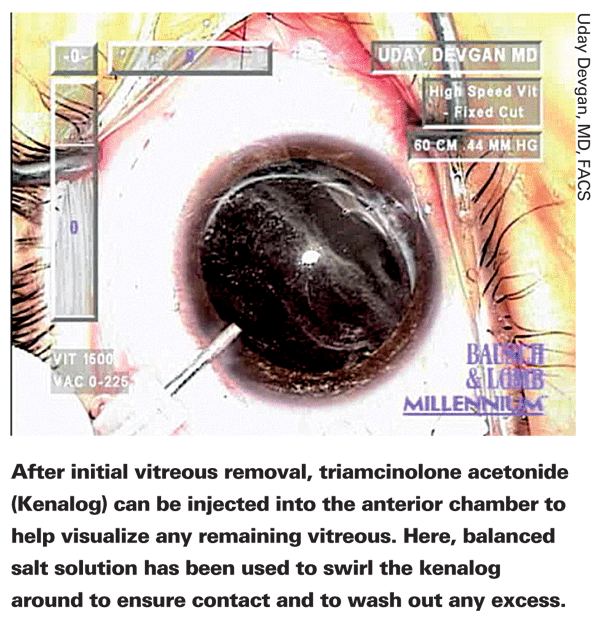
• Consider using Kenalog or air to visualize the vitreous. "Make sure you really have removed the vitreous," says Dr. Devgan. "A little Kenalog in the eye will stain the vitreous so you can get any remaining vitreous out."
Although Dr. Nichamin says he doesn't use Kenalog for this purpose, he agrees that it's a good way for surgeons to be sure all the vitreous has been removed from the anterior chamber. "Plus, having a little steroid left in the eye is not a bad thing," he notes.
"Most surgeons rinse preserved Kenalog [for this purpose] to reduce toxicity, though non-preserved forms are now available," he adds. "However, you should dilute it or it will stain the vitreous to the point where it looks like a snow storm. It's also important to be on the lookout for pulling or peaking of the iris, which is a sign of remaining vitreous strands, and to know how to sweep the anterior chamber. As a final resort, putting air into the anterior chamber will immediately allow visualization of remaining vitreous, because air and vitreous have a distinct interface that's easily recognized."
• Don't worry about leaving fragments behind. Dr. Ho says that it's important for a cataract surgeon to not be concerned if some lens material is problematic to remove.
"Leave it in the eye," he says. "Clear the vitreous from the anterior chamber; put the lens in the sulcus, if possible. Then close the eye. A retina surgeon can easily handle whatever is left—although it makes our job easier if the lens is already placed in the eye."
Finishing Up
Once the vitrectomy appears to be complete:
• Sweep the chamber. "Check for strands by using an instrument to gently sweep the anterior chamber," recommends Dr. Nichamin. "A supplementary sideport incision can be made to allow sweeping 360 degrees. If you catch a vitreous strand, you'll see it tug on the iris. Or, you can use Kenalog or an air bubble to make strands visible. Incisions need to be watertight, because a small wound leak may allow vitreous to migrate back up to a wound postoperatively."
• Use acetylcholine to bring the pupil down. "Using acetylcholine serves several purposes," notes Dr. Nichamin. "It helps the patient see better faster; it helps to lower IOP and avoid postop pressure spikes if a little viscoelastic is left in the eye; and in the complicated surgery setting, seeing a nice round symmetric pupil confirms that all vitreous is gone from the anterior chamber."
• Use a suture to guarantee water-tight closure. "This will help prevent endophthalmitis, for which the eye is now at increased risk," notes Dr. Devgan. "You don't want the wound to leak, because if the anterior chamber flattens a little vitreous may come forward again. Also, if you send the patient to see your retinal specialist, he may need to do a procedure, and he'll want to know that your incision isn't going to open up."
• Consider extra antibiotics. "You're more than 10 times as likely to have endophthalmitis once the barrier between the vitreous and the front of the eye is broken," explains Dr. Devgan. "It takes far fewer bacteria to cause infection in the vitreous than the aqueous because the aqueous is replaced every few hours, while the vitreous is thick and full of nutrients and doesn't move. So suture the incisions at the end of the case and consider giving some kind of intracameral or subconjunctival antibiotics, in addition to whatever topical drops you apply."
• Be forthright with the patient. Dr. Devgan also recommends involving patients in their postop care immediately. "Tell them: 'The good news is, we got out your cataract, and the lens implant is in place and it looks great. However, your capsule was weakened and experienced a break during the surgery. I don't think there will be any long-term effect, but just to be on the safe side, I'm going to have you see Dr. Retina. He'll make sure everything is fine.' You should say something right away, so patients know you're being honest and up-front and acting in their best interests."
After Surgery
With these patients, appropriate postop care is especially important.
• Send the patient to a retinal specialist. "If the capsule breaks, no matter what your approach, the patient should be seen by a retinal colleague in the immediate postop period," says Dr. Devgan. "I had one patient who required an anterior vitrectomy, and at the end of the case everything looked fine. Nevertheless, I sent him to a retina colleague just to be sure. The patient came back a week later and said, 'I saw Dr. Retina and he said you did a beautiful job, but I had one break in the periphery of my retina and he lasered it on the spot and said that I'd do fine.' I was thinking, 'Thank you, Lord!' If it weren't for that referral, the patient could have come back a month later with a detached retina."
• Monitor these patients extra closely. "These patients have to be monitored postop," notes Dr. Nichamin. "They're more likely to incur a pressure spike. And we treat them more aggressively with anti-inflammatories, although I usually use the same antibiotic regimen that I would for an uncomplicated patient. Depending on their clinical response, I may keep them on NSAIDs and steroids a little longer than other patients."
"Keep these patients on NSAIDs and steroids for at least six weeks," agrees Dr. Devgan. "The risk of cystoid macular edema after routine cataract surgery is very low, but when you break the capsule your risk is many times higher."


