|
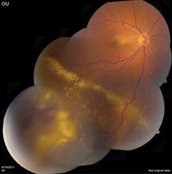
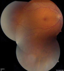
A complete peripheral retinal exam was conducted of both eyes, and revealed a yellow-red mass with extensive exudate inferotemporally in the right eye (See Figure 3). The mass was fed by a minimally dilated retinal artery and drained by a slightly dilated vein. At this point, the differential diagnosis included retinal vasoproliferative tumor and retinal hemangioblastoma. B-scan ultrasonography revealed an acoustically dense mass but did not help to narrow the differential diagnosis. A fluorescein angiogram was not performed. Given the moderately older patient age and lack of medical history, as well as the unifocality of the vascular mass and its lack of substantial vascular feeder vessels, the diagnosis of retinal vasoproliferative tumor appeared more favorable. Retinal hemangioblastoma and von Hippel-Lindau disease were felt to be less likely. The patient was treated with cryotherapy and sub-Tenon’s fascia triamcinolone (40 mg/cc) injection. At four months post-treatment, the retinal tumor showed early regression and the ERM was less prominent, with reduced macular edema and relatively stable visual acuity at 20/40. At 10 months post-treatment, the tumor was completely regressed and the ERM had retracted up to the optic disc with resolution of the retinal edema (See Figure 4). At this point, vision improved to 20/25, and it has remained stable since.
Discussion
Vasoproliferative tumor (VPT) is a vascular tumor that appears as a yellow-red peripheral retinal mass, often with related visual loss from macular edema or ERM.1 Carol Shields, MD, and Jerry Shields, MD, and colleagues initially reported a series of 12 tumors in an early 1983 paper,2 then later clarified the clinical features and management in 129 cases.1 Most recently they published a comparative update describing 334 vasoproliferative tumors in 275 patients.3 The latest publication is a comparison of the features of primary versus secondary VPT features.
|

Pathologic studies of VPT have been limited, given the rare need for enucleation or biopsy, but have consistently shown the mass to be made up of spindle-shaped cells that stain strongly positive for glial fibrillary acidic protein, which suggests a fibrocytic astrocyte cellular origin.4,5 Abnormal vascular channels with hyalinized walls are seen within the spindle cell mass. In a recent study of four cases, Lynn Janet Poole Perry, MD, and colleagues performed an array of immunohistochemical stains that showed a somewhat surprising paucity of microvascular channels despite the apparent vascularity of this tumor clinically.4 When combined with their findings of low cellular turnover and lack of markers seen in astrocytic neoplasms, the authors postulated that VPT are primarily a reactive astrocytosis associated with fibrous retinal pigment epithelial metaplasia and subretinal exudate. Marianne Smeets and colleagues likewise favor a reactive process involving both glial and vascular cells.5
In the Shields’ series, visual complaints were largely related to macular edema (24 percent), macular ERM (20 percent), macular exudate (23 percent) and vitreous hemorrhage (19 percent).3 In our case, the patient presented with mildly decreased vision secondary to macular ERM with macular edema. A wide range of retinal pathology has been implicated in ERM formation, including retinal vascular disease; inflammatory disease; post-traumatic/postoperative; retinal break; and intraocular tumors (particularly vascular tumors).6
Treatment options for VPT include observation for asymptomatic non-leaking tumors, but if there is related visual loss, subretinal fluid or exudation, then laser photocoagulation, cryotherapy, photodynamic therapy or plaque radiotherapy are considered. While no clear treatment protocol exists, our preferred treatment modality for most patients with symptomatic VPT is cryotherapy.
This case highlights the importance of a detailed peripheral retinal examination in all cases of ERM, looking for retinal breaks, inflammation and peripheral masses. The treatment of VPT is based on the individual patient’s symptoms. Observation, cryotherapy and laser photocoagulation are currently the primary treatment options.
The authors would like to thank Carol Shields, MD, and Jerry Shields, MD, of the Ocular Oncology Service at Wills Eye Institute for their invaluable assistance in preparing this case report.
1. Shields CL, Shields JA, Barrett J, De Potter P. Vasoproliferative tumors of the ocular fundus: Classification and clinical manifestations in 103 patients. Arch Ophthalmol 1995;113:615-23.
2. Shields JA, Decker WL, Sanborn GE, Augsburger JJ, Goldberg RE. Presumed acquired retinal hemangiomas. Ophthalmology 1983;90:1292-1300.
3. Shields CL, Kaliki S, Al-Daamash S, Shukla S, et al. Retinal vasoproliferative tumors. Comparative clinical features of primary versus secondary tumors in 334 cases. JAMA Ophthalmol 2013; in press.
4. Poole Perry LJ, Jakobiec FA, Zakka FR, Reichel E, Herwig MC, Perry A, Brat DJ, Grossniklaus HE. Reactive retinal astrocytic tumors (so-called vasoproliferative tumors): Histopathologic, immunohistochemical, and genetic studies of four cases. Am J Ophthalmol Pub Online Dec. 6, 2012.
5. Smeets MH, Mooy CM, Baarsma GS, et al. Histopathology of a vasoproliferative tumor of the ocular fundus. Retina 1998;18:470-2.
6. Haivala D, Parke II DW. Macular Epiretinal Membranes. In: Albert D and Miller J, eds. Albert and Jakobiec’s Principles and Practice of Ophthalmology. Canada: Elsevier, 2008:2073-82.


