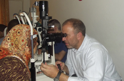
Raised in Syria, with his residency and fellowship in the United States, retina specialist Aref Rifai, MD, has been concentrating his efforts near Aleppo in the country’s northern section, and offers it as an example of the need: “Aleppo is a city of 2.5 million people and used to have about 2,000 physicians,” says Dr. Rifai. “Today, there are fewer than 100, and probably fewer than 10 ophthalmologists covering the entire city.”
Dr. Rafai is a member of the Syrian American Medical Society, and set about through SAMS and other aid groups to equip three hospitals in the rural north near the border with Turkey with ophthalmic equipment. For his first shipment, he was able to collect $70,000 to purchase a vitrectomy machine, a laser, a microscope and some ancillary equipment. Working with colleagues from the United States and the UK, Dr. Rafai now has about 10 to 12 surgeons who make week-long rotations every two or three months to supplement the efforts of local Syrian ophthalmologists.
|
SAMS is set up to accept both used medical and surgical equipment, and financial donations as well. Information on the group is available at sams-use.net. In addition to medical care, SAMS is involved in humanitarian efforts to restore decimated rural villages with the means to support themselves, such as a project to donate cows to farming areas. Another group providing relief and humanitarian services to Syria is savingfamiles.org.
Even when the immediate crisis is resolved, Dr. Rafai expects a long process before the needs of the Syrian people are met. “The infrastructure has taken a tremendous hit,” he says. “I’d estimate that 30 percent of the population is internally displaced; they don’t have a home to go to.”
Gene Variant Tied to AMD
An international team of researchers, led by scientists at the Genome Institute at Washington University School of Medicine in St. Louis and the University of Michigan School of Public Health in Ann Arbor, has identified a gene mutation linked to age-related macular degeneration.
It’s not the first gene variation linked to AMD, but it is the first to suggest a mechanism where the variant may contribute to the disease. The researchers report that a change in the C3 gene, which plays a role in inflammation and in the body’s immune response, also contributes to macular degeneration. The study was published online Sept. 15 in Nature Genetics.
“In past studies of AMD, there is a clear relationship between the complement pathway and the onset of this disease,” said co-senior investigator Elaine R. Mardis, PhD. “The complement system is part of the immune system that helps amplify or ‘complement’ the efforts of immune cells to fight infections. So the idea is that the gene variant interferes with the complement pathway’s normal function throughout life, and that can damage the retina over time, which ultimately leads to AMD’s emergence.”
The researchers sequenced DNA from 10 regions of the genome that had been linked to AMD in previous genetic studies. They analyzed a total of 57 genes in 2,335 patients with macular degeneration. Then the researchers sequenced the same genes in 789 people of the same age who did not have AMD.
The search turned up two gene variants: one in the C3 complement gene, and an alteration that had been identified in previous studies of macular degeneration.
“Finding the variant that had been identified previously helped confirm that we were on the right track,” explained Dr. Mardis, a professor of genetics and co-director of the Genome Institute. “And it’s likely this new variant was discovered because of the very large number of patients whose DNA we sequenced. By analyzing so many AMD patients, it was possible to find variants that may not have been identified in a smaller patient sample and to establish that this C3 gene variant is unique to people with AMD.”
The two gene variants together contribute to a threefold increased risk for macular degeneration. Dr. Mardis and her co-investigators hypothesize that the mutations work in tandem to increase AMD risk by interfering with the inactivation of complement in the retina.
“When you have these mutations, interactions between the proteins that cascade in the complement pathway are altered,” Mardis said. “And when they’re altered, the secondary response to infection, which involves complement, also is altered. So our hypothesis is that over time, because of the role of the complement pathway in the retina, damage begins to accrue, and eventually that leads to vision loss.”
The next step is to look at additional DNA regions in the more than 2,000 patients and controls who were involved in this study. The researchers will broaden their look across the genome and go beyond the 10 DNA regions analyzed in this study.
“We hope to identify new genes, perhaps more genes in the complement pathway, perhaps genes in other inflammatory response pathways, or in areas we wouldn’t have anticipated finding any genes related to AMD,” she said. “We’re taking a wide look at the genome to see what turns up.”
Impaired Autophagy Tied to AMD
A new study published in the PLoS One journal challenges conventional wisdom on the pathogenesis of age-related macular degeneration. The researchers found that degenerative changes and loss of vision are caused by impaired function of the lysosomal cleanup mechanism, or autophagy, in the fundus. The results open new avenues for the treatment of dry AMD, which currently lacks an efficient treatment. The University of Eastern Finland played a leading role in the study, which also involved research groups from Italy, Germany and Hungary.
AMD is a storage disease in which harmful protein accumulations develop behind the retina. These accumulations are indicative of the severity of the disease. As the disease progresses, retinal sensory cells in the central vision area are damaged, leading to loss of central vision. The cell biological mechanisms underlying protein accumulations remain largely unknown.
This is the first time that impaired lysosomal autophagy, which renders the cells in the fundus unable to dispose of old, deformed or otherwise faulty proteins, has been implicated in AMD, the researchers say. Drugs inhibiting the impairment of auto-phagy could possibly even stop the progression of AMD.
Cataract Surgery Cuts Mortality Risk
People with cataract-related vision loss who have had cataract surgery to improve their sight are living longer than those with visual impairment who chose not to have the procedure, according to an Australian cohort study published in September’s Ophthalmology. After comparing the two groups, the researchers found a 40-percent lower long-term mortality risk in those who had the surgery.
The research data was gathered in the Blue Mountains Eye Study. A total of 354 persons aged 49 years and older and diagnosed with cataract-related vision impairment – some of whom had undergone surgery and others who had not – were assessed between 1992 and 2007. Adjustments were made for age and gender as well as a number of mortality risk factors, including hypertension, diabetes, smoking, cardiovascular disease, body mass index and measures of frailty and comorbid disease. Follow-up visits took place five and 10 years after the baseline exam.
Previous research had indicated that older persons with visual impairment were likely to have greater mortality risk than their age peers with normal vision, and that cataract surgery might reduce this risk. These studies, unlike the Blue Mountains Eye Study, compared people who had undergone cataract surgery with those in the general population or with those who had not had cataract surgery, and did not link vision status to the surgical status.
“Our finding complements the previously documented associations between visual impairment and increased mortality among older persons,” said Jie Jin Wang, PhD, of the Westmead Millennium Institute and one of lead researchers of the study. “It suggests to ophthalmologists that correcting cataract patients’ visual impairment in their daily practice results in improved outcomes beyond that of the eye and vision, and has important impacts on general health.”
The association between correction of cataract-related visual impairment and reduced mortality risk is not clearly understood, but plausible factors may include improvements in physical and emotional well-being, optimism, greater confidence associated with independent living after vision improvement, as well as greater ability to comply with prescription medications.
One limitation of the study is that participants with cataract-related visual impairment who did not have cataract surgery could have had other health problems that prevented them from undergoing surgery, and that these other health problems could partly explain the poorer survival among non-surgical participants. This issue is addressed by the researchers in a subsequent study.
First Animal Model to Simulate Graves’ Disease
Researchers have developed the first animal model simulating the eye complications associated with the thyroid condition Graves’ disease, a breakthrough that could pave the way for better treatments, according to a recent study accepted for publication in the Endocrine Society’s journal Endocrinology.
Graves’ disease is an autoimmune disorder that causes the body to produce antibodies that attack the thyroid gland. The condition causes the thyroid gland to become overactive and produce too much thyroid hormone. About 1 percent of Caucasian women have autoimmune thyroid disease where the thyroid is either over- or underactive. Among those who have Graves’ disease, more than half develop eye complications, according to the study’s lead author, J. Paul Banga, PhD, of King’s College London School of Medicine in the United Kingdom. These complications include Graves’ orbitopathy, where swelling of tissue behind the eyes causes them to bulge outward. The condition can cause pain and lead to blindness.
“Current treatment options for eye complications associated with Graves’ disease are limited,” Dr. Banga said. “Better treatments are needed for Graves’ orbitopathy to reduce the risks of permanent disfigurement and social stigma. Having an animal model to test preventative treatments could lead to important advances that will ultimately benefit people with Graves’ disease.”
The condition is currently treated with steroids, which can cause undesirable side effects such as weight gain and osteoporosis.
Though researchers have previously developed animal models of Graves’ disease, these were challenging to replicate and none were able to simulate the eye problems seen in people with Graves’ disease.
To develop the new model, researchers injected mice with small, circular, double-stranded DNA molecules called plasmids. Over the course of three months, scientists used electronic pulses to ensure the DNA molecules were absorbed into the cells of each mouse. Mice that underwent this procedure developed eye problems like those seen in human patients who have Graves’ disease, while the control group of mice did not develop these complications.
“The new animal model opens the door for scientists to conduct needed mechanistic studies and identify preventative therapies to minimize this painful and debilitating condition,” Dr. Banga said. REVIEW


