In order to calculate the idealpower for an intraocular lens prior to surgery, cataract surgeons measure multiple dimensions of the eye and then plug those numbers into a formula. This protocol has resulted in a number of challenges for instrument manufacturers. First, it's created a need for greater accuracy and ease of measurement for key data such as axial length. That challenge has been met with considerable success, first by the IOLMaster (Zeiss Humphrey) about a decade ago, and more recently by the Lenstar LS900 (Haag-Streit).
Second, as our understanding of the eye and its optics has improved, the formulas used for lens power calculation have evolved and become more sophisticated; the trade-off is that they require more information about the structure and dimensions of the eye. That has challenged manufacturers to find ways to provide that extra information without too much additional time and effort.
Third, advances in the intraocular lenses themselves, including the advent of multifocal, toric and accommodating lenses, have increased the need for accuracy. And finally, the increasing prevalence of patients who have had previous refractive surgery has made the entire process more challenging. Here, three surgeons offer their perspective on the current status of biometry, and the instruments and approaches surgeons can choose to use for this purpose.
Optical Biometry
Perhaps the greatest single step forward in biometry measurement has been the switch from measuring distance using ultrasound to measuring using light. "The IOLMaster was the first commercially available optical biometer, and the ophthalmic community owes Zeiss a great debt for developing this instrument," observes Warren E. Hill, MD, FACS, medical director of East Valley Ophthalmology in Mesa, Ariz. (Dr. Hill specializes in intraocular lens power calculation.) "Optical biometry uses a 780-µm light wave that has eight to nine times the resolution of a 10-MHz sound wave. The measurement of axial length used to be one of the more problematic parts of IOL power calculations. When the IOLMaster came out, it made the measurement of axial length nearly perfect."
Dr. Hill notes that a key advantage of optical biometry is that the measurement is made to the center of the macula—as long as the patient is looking directly at the fixation light while the measurement is being taken. "That gives you the refractive axial length rather than the anatomic axial length," he notes. "Ultrasound tends to measure the anatomic axial length of the eye, unless you perform a vector A-B scan."
Given the speed at which technology evolves, it shouldn't be surprising that the much more recent Lenstar offers some advances not found in the IOLMaster. However, both instruments have strong points.
The Lenstar differs from the IOLMaster in several respects: It captures some data the IOLMaster cannot; in some cases, it takes the same measurements differently; and it organizes its data-capturing and management somewhat differently. Differences between the instruments include:
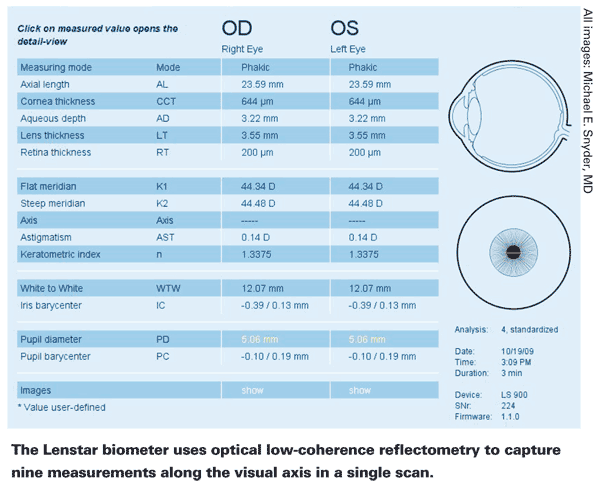
• Amount of data captured. "We've been very pleased with laser biometry, and the Lenstar has a couple of characteristics that we find very appealing," says Michael E. Snyder, MD, who practices at the Cincinnati Eye Institute in
• Measuring anterior chamber depth optically. Although both instruments measure axial length using optical biometry, the Lenstar also uses optical biometry to measure anterior chamber depth. "The latest version of IOLMaster, Version 5.4, still measures anterior chamber depth using slit imagery," Dr. Hill points out. "That measurement is very good, but it's not as accurate and reproducible as what you get using optical biometry."
In addition, the Lenstar defines anterior chamber depth somewhat differently. "Many devices measure from the front of the cornea to the front of the lens, but the aqueous depth actually is the distance from the posterior corneal surface to the front of the lens," notes Dr. Snyder. "Measuring from the posterior surface can make a difference when a patient has a fairly short anterior chamber and an unusually thick cornea.

"For example, if a cornea happens to be 700 µm instead of the average 550 µm, that adds 150 µm to the chamber depth," he continues. "Differentiating true anterior chamber depth from aqueous depth could turn out to be important; I think we may find that part of the reason our formulas don't work as well as they might is that we're not actually measuring what we think we're measuring. Hopefully, having better data will
help us to improve our formulas."
• Multiple keratometry readings. "The Lenstar takes two sets of keratometric readings: one at 1.65 mm and one at 2.3 mm," says Dr. Hill. "Taking two readings in slightly different zones and combining them via an iteration process appears to result in improved consistency."
Dr. Snyder also appreciates this feature. "The Lenstar is measuring a larger number of points," he observes. "This may give us information on the prolate or oblate nature of the central zone, which is information we haven't been able to collect before."
• Lens thickness measurement. This feature is useful when a surgeon employs one of the more recent, advanced IOL power calculation formulas. "The latest generation of lens power formulas, the Olsen and Holladay II formulas, both require lens thickness for the calculation of lens power," notes Dr. Hill. "The Lenstar gives us the lens thickness using optical biometry, and the measurement is very accurate."
• Toric IOL positioning. "I prefer laser biometry measurements to manual keratometry for selecting my astigmatism axis for implant lenses and LRIs in our multifocal patients," says Dr. Snyder. "One reason is that it captures a red-free image of the eye at the time of measurement. [See p. 48.] Currently, most people sit the patient up in the preop area and use a purple pen to mark the axis. One problem with this is that you don't know whether the patient had his head perfectly straight when the measurement was taken. A 5-degree tilt during measurement that's not there when you put the mark on will result in the implant being 5 degrees off. But if you extract this picture, you can pick a landmark and use that for alignment."
Dr. Snyder notes that the image isn't easy to extract right now. "I hope it will become easier with newer versions of the software," he says. "In fact, the Haag-Streit people have developed a slit-lamp photo that allows us to superimpose a protractor image. I'd like to see those technologies merged; then we'd know exactly which landmarks are at the axis."
• Capturing all data at once. "The Lenstar is well-thought-out, very solid technology," says Terrence P. O'Brien, MD, professor of ophthalmology and director of the Refractive Surgery Service at Bascom Palmer Eye Institute of the Palm Beaches. "One of the attractive things about it is the fact that it's an all-in-one biometer, and once the laser is properly aligned, you get all the results at once."
Dr. Snyder points out that collecting the data simultaneously is significant. "All of the measurements need to be taken along the same reference line," he notes. "If our keratometry is measured along a slightly different axis than our axial length measurement because the patient looks a little to the left or right, that can cause variability in our measurements and our final results. You can have a very accurate measurement—but it's still problematic if you're not measuring exactly what you think you are. Capturing all of these things at once will, I believe, increase our accuracy."
• Easy use of formulas. As already noted, the gathered data needs to be plugged into a formula; the steps required to accomplish that have also been an issue. The Lenstar software runs on an external PC, which allows direct communication with electronic medical record programs; the software can automatically fill in data fields in a chosen formula. Dr. O'Brien says his practice has found this to be very advantageous. "For example, the data the Lenstar measures can go directly into the Holladay IOL Consultant software," he says. "We don't have to worry about transcription errors or memory sticks going from one computer to another."
Even more exciting for some surgeons is the fact that the Lenstar can have the Holladay II formula installed onboard. "Many upper-tier surgeons use the seven-variable Holladay II formula for lens power calculation," says Dr. Hill. "That software normally has to run on a stand-alone computer. This software package has now been integrated into the biometry instrument itself, with the Lenstar EyeSuite software automatically porting the Lenstar measurements into the Holladay IOL consultant program. For example, when you bring up the Holladay II formula for a specific patient, all of the fields are filled in for you.
You don't have to use a network, or enter the data by hand. This not only saves a lot of steps, but may save some heartache in the event of a transcription error."
Dr. Hill acknowledges that many surgeons are still using the older formulas that don't require as many measurements to complete the power calculation. "The
Dr. Snyder agrees. "I can understand why some surgeons haven't switched to more advanced formulas like Holladay II," he says. "They may be thinking, 'Why should I bother switching to a different formula if I can't collect the information it requires conveniently or accurately?' I've used the Holladay II formula in some cases, but the fact that it wasn't built into the machine made it challenging. It had to be manually entered into the computer sitting at somebody's desk, and transcription is an opportunity for human error. Being able to use the newer formulas within an instrument should encourage people to use them, because it's much easier and we won't have to worry about human error."
Dr. O'Brien notes that the Lenstar has a learning curve. "As with any device, you have to spend some time becoming familiar with it, and you have to have an experienced operator," he says. "But we haven't run into any other problems with it."
IOLMaster's Strengths
"The IOLMaster and Lenstar both have strengths," notes Dr. Hill. "One concern that people had when the IOLMaster first came out was that it could only measure through cataracts up to a certain density. Beyond that point you didn't get a high enough signal-to-noise ratio for the measurement to be meaningful. To address this, the engineers at Zeiss did something quite clever. Instead of taking multiple individual measurements, checking them to make sure they're all correct and then taking the arithmetic mean, they've employed digital signal processing—the same technology used by the communication industry when the signal strength over large distances becomes extremely weak.
"That technology takes multiple measurements, perhaps 20, and looks at where there is and isn't a signal," he continues. "Where signal is present, you can use superimposition and constructive addition to amplify it so that it rises above the baseline. The same method can be used to remove background noise. In the latest generation of the IOLMaster, Zeiss uses this technology to amplify an otherwise poor signal resulting from a dense cataract, producing a much higher signal-to-noise ratio.1 Of course, Lenstar's ability to read through dense cataracts could evolve next year. Likewise, Zeiss may come out with their answer to the Lenstar next week."
Dr Hill adds that the latest version of the IOLMaster has very good keratometry. "That's due to a very fast computer," he notes. "As a result, the acquisition and processing of keratometry information is very quick and reproducible."
Is There Still Room for Ultrasound?
Given the impressive capabilities of both optical biometers, is ultrasound on the way out? Dr. Snyder says his practice keeps both immersion and contact ultrasound available for those occasions when laser biometry can't do the job. Dr. O'Brien's practice does, too. "I think there's still a place for ultrasound, especially in some abnormal eyes where you may wish to incorporate B-scan along with A-scan," says Dr. O'Brien. "However, the advantages of these multi-function optical biometers are so great that I could see some of the old contact biometry units gathering dust."
"Nowadays we're mainly using A- scan ultrasonography for the measurement of axial length when we encounter a very dense cataract that precludes the use of optical biometry," says Dr. Hill. "The disadvantage of both applanation and immersion A-scan biometry is that you can only indirectly determine where the sound beam is measuring. The operator must look for high, steeply rising spikes in the axial length display; those represent the anterior and posterior boundaries of the cornea and the anterior and posterior lens capsule. The more steeply rising the spikes, the more perpendicular the sound wave is to the acoustic interface, and the better the chance that you're measuring along the visual axis. However, the operator can't be absolutely sure that he's measuring to the center of the macula, especially if the patient has a peripapillary staphyloma.
"Nevertheless," he adds, "an immersion-ultrasound A-scan is fine for the great majority of patients with normal eye anatomy, as long as the pattern of the anterior spikes indicates
you're on-axis. But the moment you begin to have anatomic abnormalities your accuracy may be in question."
Dr. Hill says the most accurate form of ultrasound-based axial length measurement is a vector A/B-scan. "Using this method you perform a B-scan that runs through the optic nerve and the center of the macula, and a simultaneous vector A-scan," he explains.
"The reason for the increased accuracy is that you can direct the A-scan to exactly the right place, based on B-scan landmarks all lining up properly. The downside is that it takes more operator skill and the equipment is often more expensive."
Despite its potential advantages, Dr. Snyder is reluctant to rely on this approach. "This can increase the accuracy of the A-scan, but B-scan probes are big and bulky," he says. "I don't find that method to be as accurate as laser biometry, and I wouldn't rely on it to select my implant lens power unless I had no other alternative."
Perhaps the strongest point in favor of ultrasound biometry is that the current generation of optical biometers remains expensive. "For someone just starting an ophthalmology practice, this level of technology may be difficult to afford," notes Dr. Hill. "Biometry via A-scan ultrasound is far more affordable, and you can still get the measurements you need; it just takes more operator skill to ensure consistent accuracy. There's a place for everything."
Meeting the Challenge
"We all need to be looking for whatever technological advantage will allow us to increase precision and accuracy in a time of increasingly demanding refractive cataract surgery," says Dr. O'Brien. "Today, we see a lot of patients who had LASIK 10 or 15 years ago and are now reaching cataract age. That used to be a once-in-a-while thing; now it's a daily routine. In these demanding cases, having a unit that can accurately and reproducibly provide the IOL power is essential. And in busy clinical practices, a device that provides sophisticated technology in a user-friendly package is especially welcome.
"The IOLMaster and Lenstar units are both major advances over what we had in the past," he adds. "They help us get closer to the refractive target. They're both quick and user-friendly. Biometry is still challenging, though—no single device is perfect, and we still find that multiple measurements are often required to achieve the best outcomes. Nevertheless, the IOLMaster and Lenstar are both excellent instruments, and both will evolve over time."
Dr. Snyder is a consultant for Haag-Streit; Dr. Hill is a consultant for both Zeiss and Haag-Streit. Dr. O'Brien has no financial ties to any of the companies or products mentioned.
1. Hill W, Angeles R, Otani T. Evaluation of a new IOLMaster algorithm to measure axial length. J Cataract Refract Surg 2008;34:6:920-4.


