A 62-year-old black female presented to the Wills Eye ER for a two-week history of worsening right eye proptosis. She was previously diagnosed with conjunctivitis by an outside ER due to irritation in the same eye. When symptoms persisted, evaluation by an optometrist suggested a thyroid disease workup. At presentation, the patient described mild pain and blurred vision of the right eye, but denied any double vision.
Medical History
Past medical history was significant for hypertension, controlled with Atenolol and Amlodipine. Review of systems was negative for weight changes, fevers, chills or night sweats, respiratory or gastrointestinal changes or atypical arthralgias. The patient also denied any changes in hair or skin characteristics, heat or cold intolerance, as well as changes in energy level. Additionally, there was no history of smoking or personal history of malignancy. Family history was non-contributory.
Examination
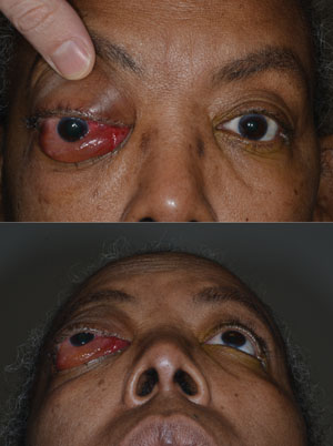
Vital signs were within normal limits. Examination demonstrated a corrected visual acuity of 20/150 in the right eye improving with pinhole to 20/70. The left eye visual acuity was 20/20. The right pupil was sluggish while the left was reactive; no afferent pupillary defect was detected and visual fields were full by confrontation. Color plates were 8/8 briskly in each eye. Extraocular muscle motility was severely restricted on the right with 90 percent deficits in all fields of gaze. Left eye motility also showed an abduction deficit of 90 percent as well as an adduction deficit of 50 percent. Prominent right eye proptosis was noted along with palpable subcutaneous nodularity. There was lower lid retraction as a result of 4+ chemosis and 3+ injection in the right eye (See Figure 1). These findings were absent in the left eye. Intraocular pressure was 22 mmHg in both eyes. Slit-lamp examination showed only superficial punctate keratopathy on the right. Dilated fundus exam was unremarkable.
|
Please click this link for diagnosis, workup, treatment and discussion.


