Presentation
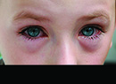
A 63-year-old Caucasian female presented to the Wills Eye Emergency Room complaining of pain radiating to her temple and ear, redness, and decreased vision in her left eye for the past week. She also noted that she had had several episodes of pain and elevated intraocular pressure in that eye over the past year, for which her ophthalmologist had performed laser surgery in each eye six months ago. She was taking timolol in both eyes once a day. She denied nausea and vomiting and her review of systems was otherwise negative.
Medical History
Her medical history was significant only for hypertension, which was well-controlled with oral medications. Family history and social history were non-contributory.
Examination
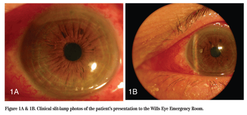
On examination the visual acuity was 20/50 OD and 20/70 OS, with no improvement with pinhole. Her pupils were round, equal and briskly reactive with no relative afferent pupillary defect. Extraocular movements were intact and confrontation visual fields were full to count fingers OU. Her intraocular pressures by applanation were 16 mmHg OD and 38 mmHg OS. Slit-lamp exam of the left eye revealed significant conjunctival injection and chemosis, mild anterior corneal stromal haze, a shallow central anterior chamber with anterior bowing of the iris, and a patent laser peripheral iridotomy. On gonioscopy, her angle was closed for 360 degrees OS, and only trabecular meshwork was visible with depression OD for 360 degrees. Both eyes demonstrated anterior bowing of the peripheral iris in a plateau-like configuration. Dilation was deferred.
Diagnosis, Workup and Treatment
The anterior bowing of the iris in the presence of a patent peripheral iridotomy led to suspicion that the patient could have angle closure secondary to a posterior segment process. A B-scan was done and revealed a severely thickened sclera, as well as a classic T-sign, representing fluid in tenon's space (Figure 2a), and anterior nasal choroidal detachments (Figure 2b). Ultrasound biomicroscopy of both eyes was performed. The right eye had a shallow anterior chamber and anterior rotation of the ciliary body causing anterior bowing of the peripheral iris consistent with plateau iris. The left eye revealed a shallow AC and a closed angle. The iris was rotated forward and the ciliary processes were against the iris pigment epithelium. Shallow choroidal effusions were seen in all quadrants.
The patient was diagnosed with acute angle-closure glaucoma secondary to posterior scleritis and ciliochoroidal effusion OS and plateau iris configuration OU. After controlling the pressure with aqueous suppression and cycloplegia, the patient was started on oral Prednisone 60 mg, Naproxen 500 mg and Ranitidine 150 mg, each p.o., b.i.d. A scleritis workup was performed, however, no associated systemic disease process was found. The patient"s pressure and scleritis both improved with oral steroids, topical aqueous suppressants and cycloplegics.
Discussion
Posterior scleritis is a granulomatous inflammation occurring either within the scleral tissue or in the adjacent vascular networks. This inflammatory process can cause fluid to leak from the choroid into the suprachoroidal space, leading to ciliochoroidal effusion. It occurs in 2 to 12 percent of all scleritis cases and occurs both in isolation and in association with anterior scleritis. Signs include proptosis, visual loss, restricted motility, choroidal folds, exudative retinal detachment, disc edema and non-pupillary block angle-closure secondary to ciliochoroidal effusions. Patients will complain of pain, both localized and referred, and severe tenderness. 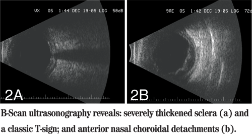
Posterior scleritis is associated with elevated IOP in 12 to 46 percent of cases, and the high pressure can be attributed to a number of causes: inflammatory debris obstructing the trabecular meshwork; inflammation of the outflow tract; peripheral anterior synechiae; neovascularization; elevated episcleral venous pressure; and use of steroids. In addition, as in our patient, ciliochoroidal effusion can cause anterior rotation of the ciliary body and iris root into the angle, leading to angle closure without pupillary block.
Angle-closure glaucoma caused by ciliochoroidal effusion can be confused with primary angle closure or malignant glaucoma. It is important to differentiate these entities because the treatments are very different. In primary angle-closure glaucoma, the central anterior chamber depth is only slightly shallow as compared with normal (2 to 2.5 mm vs. 3 mm), and it tends to be symmetrical bilaterally. The depth remains unchanged by elevated IOP and by laser iridotomy, as well. The IOP in an acute attack is usually above 50 mmHg, and responds to pilocarpine and peripheral iridotomy.
In eyes with ciliochoroidal effusion-induced angle closure, the central anterior chamber depth will be very shallow, the IOP is typically between 30 and 40 mmHg., and the attack is unresponsive to peripheral iridotomy and pilocarpine. Also, patients with primary angle closure are typically hyperopic (our patient was not); ciliochoroidal effusion can occur in emmetropes and myopes.
Malignant glaucoma is rare and is believed to be due to misdirection of aqueous flow into the posterior segment. It can occur after glaucoma filtering surgery, iridectomy, cataract extraction and with use of miotics. Its presentation is similar to ciliochoroidal effusion-induced angle closure with shallow central anterior chamber depth, flat peripheral chamber, and ineffectiveness of peripheral iridotomy and pilocarpine. However, in distinction from ciliochoroidal effusion-induced angle closure, ultrasonography and ophthalmoscopy will fail to show choroidal elevation.
Management of angle-closure secondary to ciliochoroidal effusion is directed at two processes. The first is uveal inflammation, which is treated with oral steroids to reduce the effusion and allow the chamber to deepen spontaneously as the inflammation subsides. The second is anterior rotation of the lens-iris diaphragm, which is reversed with cycloplegics. The pressure is treated in the acute setting with aqueous suppressants and oral carbonic anhydrase inhibitors. Pilocarpine is contraindicated because it will cause anterior rotation of the lens-iris diaphragm. Early diagnosis in these cases is important because it will avoid contraindicated therapy, and prompt treatment often leads to complete recovery with excellent visual prognosis.
Dr. Hoskins would like to thank William Benson, MD, and Jonathan Myers, MD, Wills Eye Institute, for their assistance.
Ikeda N, et al. Ciliochoroidal Effusion Syndrome Associated with Posterior Scleritis.
Jain SS, et al. Posterior Scleritis Presenting as Unilateral Secondary Angle-Closure Glaucoma. Indian J Ophthalmol 2004; 52(3):241-244.
Benson WE, et al. Posterior Scleritis. Surv Ophthalmol 1988;32:297-316.
Watson PG, et al. Scleritis and Episcleritis. Br J Ophthalmol 1976;60(3):163-91.
Quinlan MP, et al. Angle-Closure Glaucoma Secondary to Posterior Scleritis. Br J Ophthalmol 1978;62:330-335.
Fourman S. Angle-Closure Glaucoma Complicating Ciliochoroidal Detachment. Ophthalmology;1989;96:646-653.