
Presentation
A 67-year-old male was admitted to an outside hospital for pancytopenia and was diagnosed with acute promyelocytic leukemia (APL). He was started on chemotherapy and subsequently developed APL differentiation syndrome, a life-threatening complication of uncertain pathogenesis that includes fever and multisystem organ failure. After another two weeks, he developed a new onset of decreased visual acuity of his right eye to 20/400, periorbital edema, and right greater than left-sided retinal hemorrhages. The retinal hemorrhages were thought to be related to the patient's thrombocytopenia, and an orbital CT scan was unremarkable. His blood cultures grew vancomycin-resistant enterococcus, so he was started on broad spectrum antimicrobials. His vision subsequently worsened to no light perception within several days. A combination of orbital MRI and CT imaging now revealed preseptal soft tissue swelling with enhancement of the right optic nerve and inferior and medial rectus muscles. After normal results were obtained from a lumbar puncture, dexamethosone was started for possible idiopathic orbital inflammatory disorder. Because the patient failed to respond to these treatments, he was transferred to
Medical History
The patient's medical history included APL, Factor VII deficiency, hypertension, hypercholesterolemia and prostate cancer treated with brachytherapy. He did not have a history of diabetes. Family history was noncontributory. Socially, he admitted to a distant 60 pack-year smoking history, but denied alcohol or IV drug use. Review of systems was otherwise positive for shortness of breath, depression, fatigue, weakness and night sweats.
Examination
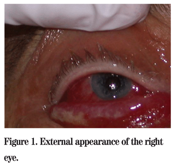
His examination revealed a visual acuity of NLP OD and 20/25 OS. His right pupil was amaurotic, and the left was briskly reactive to light. Motility testing revealed a frozen globe OD, and full motility and confrontation visual fields OS. Intraocular pressures by tonopen were 14 mmHg OD and 10 mmHg OS. External pen light exam showed 3+ conjunctival chemosis and injection OD (See Figure 1), and no abnormalities OS. Of note, he was proptotic with increased resistance to retropulsion of the right eye. Dilated funduscopic exam showed diffuse retinal whitening and optic nerve pallor OD in addition to bilateral intraretinal hemorrhages.
Diagnosis, Workup and Treatment
The patient presented with a unilateral optic neuropathy associated with chemosis, complete restriction of extraocular motility and proptosis while receiving treatment for promyelocytic leukemia. Repeat MRI of the head and orbits showed extensive signal abnormality involving the right orbit, face and pterygopalatine fossa with tenting of the right globe and signal abnormality in the anterior right temporal lobe (See Figure 2).
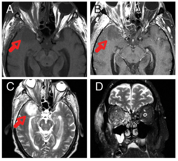
Figure 2. MRI of the orbits. A: Axial T1 image without gadolinium. B: Axial T1 image with gadolinium and fat suppression. C: Axial T2 image. D: Coronal T2 image. Note tenting of the right globe and enhancement of the right orbital contents, ethmoidal air cells and maxillary sinus. There is a collection in the right temporal lobe (arrows), which is consistent with phlegmon or early abcess formation.
The differential diagnosis includes infectious (bacterial or fungal), infiltrative (leukemic) and, though less likely in this case, inflammatory conditions (sarcoidosis, Wegener's granulomatosis, amyloidosis), or vascular causes (carotid-cavernous fistula, cavernous sinus thrombosis). In conjunction with ENT and neurosurgery, the patient was taken to the OR for orbital biopsy, exenteration and extensive debridement of necrotic tissue (See Figure 3). The sinuses and temporal lobe were also debrided. Based on the pathology results, the diagnosis of rhino-orbito-cerebral mucormycosis was made. The patient was started on liposomal amphotericin, underwent several more debridement surgeries and was discharged to a rehab facility in stable condition after almost four months of treatment.
Discussion
Rhino-orbito-cerebral mucormycosis (ROCM) is a rare, fulminant opportunistic fungal infection that is mostly seen in immunocompromised or diabetic patients. Mucormycosis refers to infection due to fungi in the order of Mucorales, with Rhizopus species being the most common causative organism. These fungi are ubiquitous and are commonly found in soil, decaying matter or moldy bread. Due to the low virulence of these organisms, disease mainly occurs in hosts who are immunocompromised with poorly controlled diabetes mellitus (especially with ketoacidosis), chronic steroid use, hematologic or solid malignancy, neutropenia or burns.
When spores are deposited in the nasal turbinates, they transform into fungal hyphae which invade blood vessels, producing tissue thrombosis, infarction and necrosis. ROCM classically begins as sino-orbital disease, presenting with a unilateral headache, facial pain, fevers and nasal congestion. Late-state disease may progress to progressive facial cellulitis, black nasal discharge or eschar of the palate or nasal septum. Later, the rhino-orbital state ensues with orbital signs and visual loss due to orbital apex syndrome and/or ophthalmic artery occlusion. The final stage of rhino-orbital-cerebral disease presents with focal neurologic symptoms and decreased consciousness.
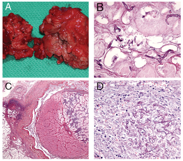
Figure 3. A: Exenterated orbital contents. Note the dark ischemic orbital tissue. B: H&E section of orbital contents shows multiple nonseptate branching hyphae consistent with the diagnosis of mucomycosis; C: H&E section shows hemi-infarction of optic nerve. D: H&E sections shows numerous hyphae obstructing the central retinal artery.
A high index of suspicion is fundamental to making this life-threatening diagnosis in a timely manner. Head and facial MRI and CT scans are the modalities of choice to evaluate orbital disease and to provide information for planning the extent of surgical debridement. However, it is imperative to recall that radiographic findings usually lag behind clinical progression in this disease, and a negative initial imaging study does not provide a rationale to delay more aggressive diagnostic maneuvers if clinical suspicion is high. The definitive diagnosis of mucormycosis is established by obtaining a biopsy specimen of the involved tissue. Stains with hematoxylin and eosin (H&E), Grocott methenamine-silver (GMS) or periodic acid-Schiff (PAS) demonstrate broad, irregular, nonseptate, right-angled, branching hyphae. Vascular invasion and necrosis as well as a neutrophil infiltrate is typical.
The mainstay of treatment for mucormycosis is correction of the underlying systemic abnormality, antifungal therapy and early surgical debridement. In light of antimicrobial resistance, a combination of liposomal or IV amphotericin B, an azole and/or caspofungin is usually initiated. In conjunction with ENT and neurosurgery, emergent sinus drainage and debridement of infected orbital or brain tissue is essential. Repeated debridement of involved tissue is often necessary as dictated by serial CT or MRI surveillance.
Mucormycosis has an extremely high mortality rate even with aggressive surgical intervention. The mortality rate associated with rhinocerebral disease is 50 to 70 percent, depending on the underlying immune problem. Additionally, rhinocerebral disease causes significant morbidity in patients who survive because treatment usually requires extensive, and often disfiguring, facial surgery. However, if the correct diagnosis is promptly made, a full clinical recovery may be achieved.
Dr. Jadico would like to thank Robert Penne, MD, of the Oculoplastic and Orbital Surgery Service at Wills Eye Institute, and Ralph Eagle, MD, of the Wills Pathology Service, for their assistance with this case.
Dhiwakar MA, Thakar A, Bahadur S. Improving outcomes in rhinocerebral mucormycosis-—early diagnostic pointers and prognostic factors. J Laryngol Otol 2003;117:861-865.
Nithyanandam S, Jacob MS, Battu RR, Thomas RK, Correa MA, D'Souza O. Rhino-orbito-cerebral mucormycosis. A retrospective analysis of clinical features and treatment outcomes. Indian J. Ophthalmol 2003;51:231-236.
Munir N,
Spellberg B, Edwards J, Ibrahim A. Novel perspectives on mucormycosis: pathophysiology, presentation, and management. Clin Microbio Rev 2005 Jul;18(3):556-569.
Talmi YP, Goldschmied-Reouven A, Bakon M, Barshack I, Wolf M, Horowitz Z, Berkowicz M, Keller N, Kronenberg J. Rhino-orbital and rhino-orbito-cerebral mucormycosis. Otolaryngol Head Neck Surg 2002 Jul;127:22-31.


