Presentation
A 32-year-old white female with a history of migraines presented to the Wills Eye Neuro-ophthalmology Service with a two-week history of a constant headache and three different episodes involving her vision. Two weeks earlier the patient described the first visual episode as "seeing strobe lights" in both eyes lasting for 30 minutes. This episode later evolved into a constant migraine headache. She described her baseline migraines as common migraines with a retrobulbar, dull pressure-like headache accompanied by occasional photophobia and nausea without visual scintillations, occurring once per month.
Three days later the patient experienced difficulty focusing in which she "couldn't see text messages," associated with "difficulty with depth perception," dysarthria, and right-sided weakness of her face and hand lasting for approximately 20 minutes. At this time, the patient was seen at an outside emergency room and discharged home after a normal CT scan of her head.
The patient's third visual episode occurred the following day as intermittent periods of right homonymous hemianopsia lasting a few seconds each time. Her symptoms resolved after approximately 20 minutes and were associated with posterior neck pains.
At the time of presentation to the neuro-ophthalmology service, the patient denied visual symptoms. There was no associated diplopia, dysphagia, gait instability or trauma.
Medical History
The patient had a medical history of migraine headaches diagnosed 10 years ago by a headache specialist. She did not have any history of diabetes, hypertension or hypercholesterolemia. Her family history and review of systems were noncontributory. The patient had a history of smoking three cigarettes a month for the past 10 years and denied alcohol and intravenous drug abuse.
Examination
The patient's examination revealed a corrected visual acuity of 20/20-2 in the right eye and 20/20-2 in the left eye. Color plates were eight out of eight briskly in each eye with normal Amsler grids bilaterally. Gross exam showed no mass, proptosis, lid lag or lid retraction. Slit-lamp exam was white and quiet. Intraocular pressure was 15 mmHg in her right eye and 12 mmHg in her left eye. Pupils were briskly reactive without an afferent pupillary defect or Horner's syndrome. The patient was orthophoric with full ductions and versions without diplopia or nystagmus. Visual fields were full by confrontation and by Humphrey perimeter. Dilated fundus exam was normal.
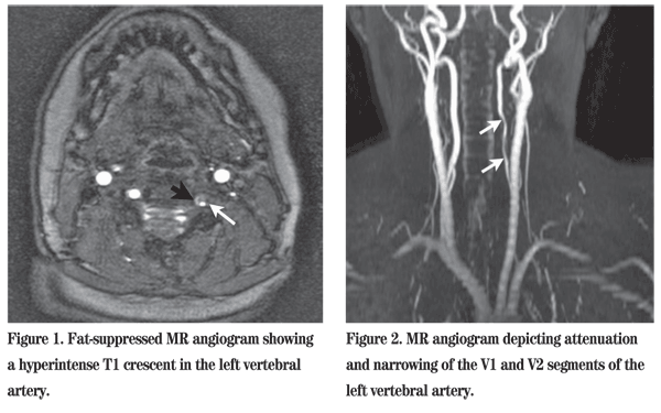
Diagnosis, Workup and Treatment
Discussion
Headache is often the most common initial manifestation of symptomatic vertebral artery dissection. In a study performed at the Mayo Clinic, headaches in vertebral artery dissections were often described as gradual in onset, unilateral and localized to the occiput, ipsilateral to the side of dissection. However, headaches in vertebral artery dissections have occasionally been characterized as sudden onset, bilateral or generalized. Neck pain is a hallmark feature of vertebral artery dissection and is more common in vertebral artery dissection than carotid artery dissection. Lateral medullary syndrome is often associated with headache and/or neck pain. A 1994 study found that ocular manifestations of vertebral artery dissections included diplopia, blurred vision, transient visual obscurations, nystagmus, Horner syndrome, decreased corneal sensation and visual field defects. A case report by Glen Jickling, MD, and colleagues describes a left vertebral artery dissection causing bilateral internuclear ophthalmoplegia. Risk factors for vertebral artery dissections include spinal manipulation, minor neck trauma, yoga, judo, ceiling painting and fibromuscular dysplasia.
Conventional angiography remains the gold standard of cervical artery dissections. However, conventional angiography is invasive and does not depict arterial wall thickness or allow simultaneous representation of the surrounding vessels in the brain. In a prospective study, Corinne Levy, MD, et al. found that MR angiography, with 95-percent sensitivity and 99-percent specificity, was more accurate in detecting carotid artery dissections than MR imaging, with 84-percent sensitivity and 99-percent specificity. However, these results were not statistically significant.
Alternatively, they found the sensitivity to be 20 percent and the specificity to be 100 percent in vertebral artery dissection for MR angiography, while sensitivity was 60 percent and specificity 98 percent for MR imaging. The authors suggest the variable caliber of the vertebral arteries in conjunction with their small diameter as reasons that MR angiography fails to detect vertebral artery dissections. James M. Provenzale, MD, proposes that conventional angiography be performed in any patient with a high clinical suspicion of a vertebral artery dissection and a normal or indeterminate MR.
Alternatively, the role of CT imaging and CT angiography seems promising. In a retrospective study of 18 patients with 25 cervical artery dissections,
Treatment for vertebral artery dissections has not been widely studied. Most treatment regimens involve anticoagulation for at least three to six months in cases without intracranial hemorrhage. MR imaging and MR angiography provide a non-invasive means for follow-up to ensure patency of the narrowed or occluded arterial lumen. If patency is reestablished, anticoagulation is often followed by antiplatelet therapy indefinitely. Marcel Arnold, MD, and colleagues examined predictors of outcomes in 169 patients with vertebral artery dissections. They found that vertebral artery dissections most commonly occur in the V2 and V3 segment and found that a low National Institute of Health stroke scale score and young age are independent predictors of a favorable outcome.
Dr. Pao would like to thank Robert Sergott, MD, and Deepak Grover, DO, of the Wills Eye Institute Neuro-Ophthalmology Service, for their time and assistance with this case.
1. Arnold M, Bousser MG, Fahrni G, et al. Vertebral artery dissection: Presenting findings and predictors of outcomes. Stroke 2006;37:2499-503.
2. Hicks PA, Leavitt JA, Mokri B. Ophthalmic manifestations of vertebral artery dissection: Patients seen at the mayo clinic from 1976 to 1992. Ophthalmology 1994;101:1786-92.
3. Jickling G, Leung K, Gan K, et al. Left vertebral artery dissection causing bilateral internuclear ophthalmoplegia. CJEM 2008;10:485-7.
4. Levy C, Laissy JP, Raveau V, et al. Carotid and vertebral artery dissections: Three-dimensional time-of-flight MR angiography and MR imaging versus conventional angiography. Radiology 1994;190:97-103.
5. Provenzale JM. Dissection of the internal carotid and vertebral arteries: Imaging features. AJR Am J Roentgenol. 1995 Nov;165(5):1099-104.
6. Schievink WI, Mokri B, Whisnant JP. Internal carotid artery dissection in a community: Rochester, Minnesota, 1987-1992. Stroke 1993;24:1678-80.
8. Vertinsky AT, Schwartz NE, Fischbein NJ, et al. Comparison of multidetector CT angiography and MR imaging of cervical artery dissection. Am J Neuroradiol 2008:29:1753-60.


