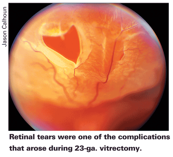
Patients with (or suspect for) glaucoma, as well as those with a gas fill, may be at risk for high postoperative intraocular pressure during the first month following 23-ga. pars plana vitrecomy. This new data comes from a retrospective review including all eyes undergoing the procedure with at least one-month follow-up at the Wayne State University Department of Ophthalmology in

Results showed that IOP spikes >22 mmHg in the first month occurred in 73 percent of eyes presenting with (or suspect for) glaucoma, versus 46 percent of eyes without glaucoma (p=0.017); 76 percent of eyes with a gas fill versus 44 percent of eyes with a fluid fill (p=0.0036); and 21 percent of eyes started on IOP-lowering drops on postoperative day one versus 49 percent of eyes that were not (p=0.0033). Complications included retinal tears (3 percent), intraoperative retinal detachment (2 percent) and postoperative retinal detachment (2 percent). Fifteen percent of eyes required suturing of at least one sclerotomy. There were no cases of postoperative hypotony or endophthalmitis. The authors conclude that aggressive early treatment of IOP may prevent spikes in the early postoperative period.
Retina 2010;30:629-34
Singh CN, Iezzi R, Mahmoud TH.
Bioceramics Orbital Implants Shown to Have Postoperative Exposures
A study at the
There were 353 patients followed for three to 96 months with an average of 30 months of follow-up (median 23 months). The authors found that implant exposure had occurred in 32 of those patients (9.1 percent). Six of the 32 exposures (19 percent) occurred during the 90-day postoperative period (average 2.1 months). The remaining 26 exposures (81 percent) occurred outside of the 90-day postoperative period (average 27.5 months, range four to 82 months).
Ophthal Plast Reconstr Surg 2010;26:80-2
Glaucoma Patients Present Mostly Error-Free OCT Scans
New research has shown that most glaucoma patients can produce artifact-free optical coherence tomography scans. In an effort to demonstrate the types and prevalence of errors associated with the use of OCT in a cross section of glaucoma patients, researchers at the Duke University Eye Center in Durham, N.C., evaluated OCT data from glaucoma patients in a three-month period for evidence of errors in peripapillary retinal nerve fiber layer maps and the macular thickness maps. Signal strengths, centering errors, individual scan errors and association with ocular conditions were noted. Logistic regression was used to assess the significance of continuous and categorical variables in predicting the presence of artifacts.
The authors observed that macular scan artifacts were present in 16.8 percent, and RNFL scan artifacts in 15.7 percent, of 89 eyes of 89 patients studied. RNFL off-center scan was the most common error (34.8 percent). For macular thickness, 100 percent of the scans were artifact-free for signal strength 8 or higher. However, for signal strength ¡Ü4, 64.3 percent had artifacts. For RNFL thickness, with a signal strength ¡Ý6, 96 percent of the scans had no artifacts. However, for signal strength ¡Ü4, 86 percent had artifacts. Macular scan artifacts were present more commonly in patients with dry eye, and RNFL centering errors were more frequent in eyes with cataracts.
J Glaucoma 2010;19:237-42
Asrani S, Edghill B, Gupta Y, Meerhoff G.


