Here, Dr. Osher discusses three new developments in the field of cataract surgery that may help to make this operation easier and safer. Two of them were designed by Dr. Osher himself; the other is a device that he discovered in Europe and is eager to introduce to American surgeons. (Dr. Osher has no financial interest in any of these instruments.)
A Better Surgical Drape
Dr. Osher has a long history of being interested in the sterile field and the design of surgical drapes; he designed the first split-lid drapes, the first sterile-lid drapes and the first drape to cover the phaco machine, allowing the scrub tech to adjust the machine instead of the circulating nurse.
“There have still been a lot of problems with the standard set of drapes,” Dr. Osher notes. “Patients would complain, ‘It’s really hot under here.’ They’d say, ‘I’m claustrophobic.’ Or they’d say to the nurse, ‘Please hold my hand,’ but there was no way the nurse could get to the patient’s hand. The drape would sometimes hang down over the patient’s head so far that it would be lying on the phaco pedal; when I’d press on the pedal, the drape would turn the patient’s head. The long drape also made it difficult for the anesthesiologist to get to the IV. In addition, I didn’t like the cardboard feel of the current drapes; they weren’t pliable or easy to work with. And patients would tell me that the worst part of the cataract surgery was when I removed the drape at the end, because of the strong adhesive. I was disturbed to see how red and irritated patients’ skin would be as a result of removing the drape.
“I felt we should be able to do better,” Dr. Osher says. “So
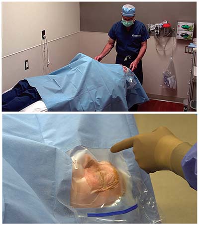 |
| The Visidrape Osher Half Body Aperture Drape is shorter than standard drapes, to improve patient comfort and surgical team access; the new material is more comfortable and less hot for the patient; the adhesive has been modified to eliminate patient discomfort when the drape is removed; and the new design allows some light to reach the unoperated eye, making patients less claustrophobic. |
“From the surgeon’s perspective, it’s a universal drape design, so it’s good for the left or right eye,” he continues. “It forms an inverted-D shape, so it’s parallel to the brow and runs along the bridge of the nose. It comes down on the lower lid and then up along the temple on either eye, so it gives you beautiful exposure while at the same time acting as an effective barrier. Also, the length of the drape had been shortened to allow the anesthesiologist and nurses easy access to the patient, while making it less hot underneath for the patient and preventing the drape from catching on the foot pedal.”
Dr. Osher says that the surgeons at the Cincinnati Eye Institute are now beginning to use the new drape. “I still use my split-lid drapes to cover the lashes,” he notes. “They’re enclosed in the package. I also continue to use a little wick to remove fluid from the lacrimal lake, and I still cover any exposed skin with a steri-strip on the lateral canthal angle. I’m very meticulous about draping, and I’m enthusiastic about having this new drape design.”
The Visidrape Osher Half Body Aperture Drape is sold in shelf packs of 10 drapes and is available from Beaver-Visitec International (Waltham, Mass.) You can find more information in the product catalogue at beaver-visitec.com, or by calling BVI customer service (866-906-8080) or your local BVI sales representative.
Sterilizing the Air
The Operio Mobile is a new instrument made by Toul Meditech in Sweden that’s designed to sterilize the air around the operating field by creating a directed, laminar, nonturbulent, ultraclean air flow over the surgical site and/or sterile instruments. The unit
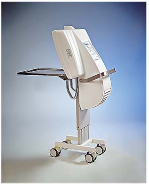 |
| The Operio Mobile, from Toul Meditech in Sweden, creates a directed, laminar, nonturbulent, ultraclean air flow over the surgical site, removing bacteria and particles from the air, to help ensure that the surgical site remains sterile throughout the procedure. |
Dr. Osher, who has been using the first Operio instrument in the United States, discovered the technology at the 2016 European Society of Cataract and Refractive Surgeons meeting in Copenhagen. “We’d had several experiences at the Cincinnati Eye Institute that were of great concern to me,” he says. “The surgical staff noticed an occasional piece of lint falling on the surgical field. Because most of the fibers were blue, we suspected they were coming from the surgical drapes.
“Then, right before attending the ESCRS meeting, one of my patients developed a corneal abscess after a fiber got caught in the stab incision,” he continues. “We had to take the patient back to surgery, remove the fiber and give intracameral vancomycin. Fortunately, the patient did fine—but I didn’t! It was traumatic for me; I’d never had anything like that happen in my career. It taught me that all airborne particulate matter is not benign, which is what I’d always thought.
“That’s when I discovered the Operio at ESCRS,” he says. “The model I saw is a mobile device that’s very easy to use; it’s like pulling up a monitor next to the eye. After we adjust the microscope and move the phaco machine closer to the patient, Operio is rolled into position either above the patient’s head for a right eye, or on the right side of the patient’s head for a left eye. It generates a laminar flow that sterilizes the field wherever it’s pointing.
“I was so excited about the technology that they allowed me to bring a mobile unit back to the United States,” he continues. “Operio was in the process of receiving FDA clearance, so I jumped at the chance to try the technology. Since then, we’ve been using it in practice, and we’re also conducting a clinical study to see how effective the device actually is.”
Dr. Osher says he’s seen two remarkable things. “First, we’ve observed a total absence of particulate such as lint in the operative field since we began using Operio,” he says. “Second, our study has found a dramatic reduction in the objective counts of 5-µm-size particles in the air—the size of bacteria. That’s significant, because there have been plenty of articles over the years demonstrating that there are bacteria in the air. I remember a study by Stuart Brown, MD, years ago, which found bacteria growing on culture plates that were left open in the operating room.”
Julia G. Schneider, MD, senior resident in ophthalmology at the University of Cincinnati Medical Center and Cincinnati Eye Institute, designed the study and is working on it with Dr. Osher. “We developed an in-vivo protocol designed to compare the presence of particulate matter ranging from 0.3 to 5 µm in size that are present in the operating room, with or without the Operio present,” she explains. “This was done by using a specialized particle counter. We take readings in the OR outside the range of action of the Operio; within the sterile field prior to turning on the Operio; and finally within the sterile field after turning on the Operio. Patients are prepped and draped in the standard fashion and the surgery proceeds as usual.
“So far we’ve looked at 75 cataract procedures utilizing our protocol,” she says. “The results have been very impressive. In all cases there’s been a vast reduction in particulate matter measured within the sterile field after utilizing the Operio. In some cases, the particles of the measured size have been completely expunged.” Dr. Schneider says they hope to have the study completed in October and publish it within the next six months.
Dr. Osher says he believes a tool like this could have implications for other areas of ophthalmology besides anterior segment surgery. “I’m especially
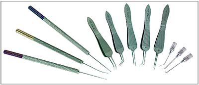 |
| The new No Fly surgical tools from Bausch + Lomb are designed to keep the surgeon from accidentally striking low-hanging instruments such as aberrometers. The line includes (from left to right): a Y-Hook; chopper; manipulator; tying forceps; capsule/foreign body forceps; fixation forceps; incision forceps; suturing forceps; a hydrodissection cannula; dye cannula; and a J cannula 2.0. |
“To me, deciding to use this instrument was a no-brainer,” he concludes. “It’s true that I only encountered one case of corneal abscess due to a fiber, but having now used the Operio many times and seeing the early results of our study, I’m a believer. I use it in every one of my cases, and it gives me a great sense of security. It makes me feel that I’m creating the safest possible field in which to perform surgery.” For more information, contact Fran Carleton at Vitreq USA by phone (603) 347-5590 or email (csc@vitrequsa.com).
Helping Keep Handheld Tools Sterile
Dr. Osher says he developed the new Storz Ophthalmics line of No Fly surgical tools (Bausch + Lomb) in response to the evolution of devices that are placed under the operating microscope, close to the surgical field. “We’ve developed all kinds of devices that hang down from the microscope, including aberrometers, the Verion interface and the Callisto interface,” he points out. “As these devices started to encroach upon the surgical field, I found myself occasionally clunking my handheld surgical tools against them, compromising their sterility. It didn’t take a rocket scientist to think of making the instruments shorter so they wouldn’t stick up and hit the microscope add-ons.
“There is a very brief learning curve to using the new tools, because you use fingertip control a little bit more,” he admits. “But other than that, I don’t see any downside to using these instead of the standard handheld instruments. I use them all the time, even if nothing is hanging down below the microscope.”
The No Fly instruments currently available from Storz Ophthalmics include five types of No Fly forceps (tying, incision, fixation, capsule/foreign body, suturing), as well as a No Fly chopper, No Fly manipulator and No Fly Y-hook. (For more information, visit storzeye.com.) REVIEW


