The Diagnostic Pyramid
In an effort to properly diagnose and subsequently treat refractive surgery patients with tear-film abnormalities, I codified my thought process into a decision pyramid. The diagnosis methods begin at the top with the more straightforward, routinely used ones and eventually lead downward toward the more involved methods for patients whose problems are harder to pinpoint. I try to turn the subjective symptoms into an objective diagnosis, thus determining whether a patient has dry eye or another condition. If a patient is pigeonholed as having dry eye when he actually is suffering from something else, he could be relegated to years of using the wrong drops without relief.
• First level: Routine tests. After listening to the patient describe his symptoms, the first step toward determining their cause consists of in-office tests such as Schirmer"s tear test, fluorescein, rose bengal and lissamine green staining, and tear-film clearance and breakup time.
You can also gather important information by examining the tear film at the slit lamp with the room light dimmed. You will see the tear lake and may see excessive tear splashing in the ocular fornix, which will mean a deficiency in the oil layer in the tear film. In this case, the patient may not have aqueous-deficient dry eye, and instead may be suffering from meibomitis. Artificial tears won"t solve the problem. You can instead advise lid scrubs, warm compresses or oral doxycycline. A liposomal spray may be helpful in such conditions.
Also, find out if the symptoms get worse or better during the day. If they"re worse in the morning, it"s probably meibomitis, not dry eye. This is because as the day progresses the eye washes the toxic tears away. If it were dry eye, however, it would be the opposite: The eyes would be comfortable in the morning after having had the lids closed all night, but then would become drier during the day as they remain open. If the patient has itching, this is a key sign of allergy; obviously, bulbar edema and papillae in the upper lid clinch the diagnosis.
• Second level: Tear-film analysis. If you"re still not certain what is causing a LASIK patient"s complaints of dryness and grittiness after performing the usual in-office tests, you can perform tear-film analysis and measure lactoferrin and IgE levels and tear osmolarity. If it is dry eye, a lactoferrin microassay with a device such as the Corneal Science Tear Profile Analyzer (Touch Scientific, Raleigh, N.C.) will determine whether it is aqueous-deficient dry eye (lactoferrin levels below 0.9 mg/ml) or evaporative dry eye (lactoferrin levels greater than 1.8 mg/ml). IgE levels will indicate ocular allergies, and if they"re high, the patient will need anti-allergy medication, rather than artificial tears. In the near future, gram negative and gram positive testing will also be possible with this equipment.
This testing can be a great help in determining the appropriate treatment. For example, say a patient has meibomitis, but she is diagnosed as having dry eye. When the artificial tears don"t work, the clinician will naturally proceed to punctal plugs—just the opposite of what the patient needs. Now, instead of having open puncta to allow the toxic tears to flow away from the ocular surface, the toxins will stay there and could cause surface damage.
The cost of the machine ranges from $12,000 to $13,000. I have used this system and believe it has the potential to specify where in the dry-eye disease spectrum the patient lies. Though the device"s cost can be prohibitive, some practices get around the expense by sending their patients" lactoferrin samples to a central hub (usually a hospital or large practice with the machine) for testing.
• Third level: Tissue sampling. Brush and impression cytology can show the stages of ocular surface involvemement by detecting keratinization, changes in cell morphology, loss of goblet cells and other indicators. This might help determine the severity of the condition and plan a surgery such as a stem cell graft with amniotic membrane transplantation.
Next in terms of invasiveness come biopsies of the lacrimal and/or salivary glands. These could be useful in Sjˆgren"s syndrome, in which inflammatory, degenerative and infiltrative changes will be evident. They can also reveal a more serious systemic disorder for which dry eye may have just been a presenting symptom. Blood or serum tests might be necessary to confirm this.
The patient you"re evaluating for LASIK or examining postop could have classic dry eye, LASIK-induced dry eye or something else. By eliminating any false diagnoses, you can begin to treat him effectively.
Dr. Gulani is chief of the cornea and external disease department and director of refractive surgery at the University of Florida, Jacksonville. With post-LASIK dry eye emerging as the most common complication of the procedure, we have to take it very seriously, and share our integrated approaches to treating it. Only by doing this will we be able to progress toward the "super vision" that LASIK may someday provide patients.
Post-LASIK Dry Eye
If a patient presents post-LASIK with complaints of dryness, there"s a good chance it"s the temporary post-LASIK condition that mimics dry eye. Among the reasons we know of for this condition is the neurotrophic keratitis identified by Rockville Center, N.Y., surgeon Eric Donnenfeld and others. They"ve pointed out that making the corneal flap in LASIK, especially if the hinge is superiorly located, can sever the corneal nerves, resulting in a temporary neurotrophic keratitis. 
A dry spot and yellow/orange iron deposits in a patient with post-LASIK dry eye.
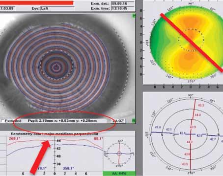
Another important reason, I believe, for dry eye post-LASIK is the change in corneal curvature between the treated and non-treated zones caused by the ablation. This creates an abrupt change in corneal curvature. This change results in the tear film having difficulty in properly draping the cornea, thereby resulting in stagnation of the tears with iron deposition forming a ring that very much resembles the deposits we see in keratoconus, the Stocker"s line in pterygium and the Ferry"s line associated with a filtering bleb in trabeculectomy. I refer to this as the pseudo-Fleischer"s ring, and it points toward the cause of dry eye in the patient.