The 3D Digital Microsurgical Workstation System from 3D Vision Systems is a 10- to 15-pound cylinder 12 inches long and 6 inches wide that is situated above the patient. The system produces two images on an accompanying wide-screen, high-definition monitor. The two images have 90-degree polarization, which, when viewed with cross-polarized 3-D glasses, yields an image that the company says has a better depth and three-dimensionality than that seen through a microscope.
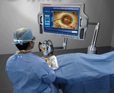 |
| A concept computer drawing illustrating how the company hopes to be able to design the system for its eventual release. |
"I was skeptical when I realized I'd have to wear special glasses," says Irvine, Calif., surgeon Roger Steinert, who consults for the company. He's used the system for several simulated surgeries and on one live patient at Arturo Chayet, MD's practice in Tijuana, Mexico. "The image is so clear and bright, though, that you don't realize you're looking through polarized glasses. Also, the system frees the surgeon from being harnessed to the microscope. You can position yourself comfortably and move your head around."
Dr. Steinert says the 3-D perspective can be beneficial. "Anytime you're doing procedures that involve looking at structures that are in the same plane as the iris, you have trouble with the regular microscope, because it uses flat illumination that doesn't distinguish different levels in the anterior to posterior direction. The place you see this most commonly is with the capsulorhexis. It's common for even experienced surgeons to grasp for the edge of the rhexis and find that it's just not where they think it is relative to where the forceps appear to be. Many things would be enhanced by the improved stereopsis, such as suturing an iris defect or epiretinal membrane peeling."
As with most new technologies, the system is a work in progress, though it's nearing a marketable stage. It may be ready for purchase in about six months, according to the company.
"In the earlier versions, if you moved the instruments quickly you could get ahead of the image processor," says Dr. Steinert. "But the last version I saw was fast enough so that there was no lag. Resolution was a concern, but it's now high-definition, so you don't perceive that you're looking at an electronic image."
But can it pry surgeons away from the microscopes they've used successfully for years?
3D Vision Systems' CEO Ram Rao thinks so. "It's more ergonomic; they don't have to look through the microscope the whole time," he says. "We also plan for it to have intelligence applications as well, such as using the system's two cameras to digitally triangulate the center of the eye to enhance intraocular lens centration. If it can help people do surgery a little better or faster, I think people would like that."


