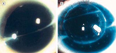
During this study to calculate clinical guidelines for optimal location and size of a rotational autokeratoplasty, researchers calculated the ideal graft size and trephine decentration for a rotational autograft based on scar location using geometric models. The model was used in a rotational auto-keratoplasty of a patient with a history of a corneal scar and diplopia. An 8-mm autograft was decentered 0.5 mm superiorly and rotated 180 degrees to relocate the scar to the superior aspect of the cornea, out of the patient's vision.
In cases that satisfy the given variables, the previously mentioned graft balances maximization of scar removal and scar movement superiorly, with minimization of discrepancy in corneal thickness after rotation, the researchers say. This resolves the diplopia and improves the visual acuity.
 |
| Preop (left) and postop (right) photographs of a 21-year-old male patient with a corneal scar, who underwent ipsilateral rotational autokeratoplasty. Copyright 2006, American Medical Association, All Rights Reserved. |
The study has important clinical implications, the researchers say, since ipsilateral rotational autokeratoplasty is not a widely used technique because of limited indications and lack of clear and easily understandable guidelines. With this study, the researchers hope ipsilateral rotational autokeratoplasty can be performed more often, as it has many advantages over keratoplasty using a donor cornea.
(Arch Ophthalmol. 2006;124:410-413)
Afshari NA, Duncan SM, Tanhehco TY, Azar DT.
Dry Eye Common Occurrence After Myopic LASIK Surgery
Dry eye is a common occurrence post-LASIK surgery, even among patients with no history of dry eye, according to a recent study. Researchers found the risk of developing dry eye correlates with the degree of preoperative myopia and the depth of laser treatment.
The single-center, prospective clinical trial included 35 adult patients, who ranged in age from 24 to 54 years, with myopia who were to undergo LASIK. The participants were randomized to undergo LASIK with a superior or nasal hinge flap. The criteria for dry eye used in the study was a total corneal fluorescein staining score >3. Visual acuity, ocular surface parameters and corneal sensitivity were also analyzed. Cox proportional-hazard regression was used to assess rate rations (RRs) with 95-percent confidence intervals. Bilateral LASIK was performed with either a superior-hinge Hansa-tome (Bausch & Lomb) microkeratome (n=17) or a nasal-hinge Amadeus (Advanced Medical Optics) microkeratome (n=18).
The patients were evaluated at one week and at one, three and six months after the surgery. The incidence of dry eye in the nasal- and superior-hinge group was eight of 17 and nine of 17 at one week, seven of 18 and seven of 17 at one month, four of 16 and three of 17 at three months, and two of 16 and six of 17 at six months, respectively. Dry eye was associated with level of preoperative myopia (RR 0.88/each diopter, p=0.04), laser-calculated ablation depth (RR 1.01/µm, p=0.01), and combined ablation depth and flap thickness (RR 1.01/µm, p=0.01).
The researchers recommend pa-tients be counseled about the risk of developing dry eye after LASIK, particularly those with high myopia. Con-sideration should be given to performing surface ablation (photorefractive keratectomy) in these patients because that technique carries a much lower risk for inducing dry eye.
(AM J Ophthalmol 2006;141:438-445)
De Paiva CS, Chen Z, Koch DD, Hamill MB, Manuel FK, Hassan SS, Wilhelmus KR, Pflugfelder SC.
Fungal Keratitis Uncommon In Northeast United States
A review of clinical and microbiology records at the New York Eye and Ear Infirmary identified 61 cases of fungal keratitis in 57 patients between January 1987 and June 2003. Fungal keratitis, the re-searchers say, is an uncommon cause of infectious keratitis in the northeastern United States.
Medical records of all patients were retrospectively reviewed to better delineate patient demographics, risk factors, etiologic organisms, treatment, and outcomes during the study to review clinical experience. A total of 5,083 positive corneal cultures were recorded during the 16-year period. Sixty-one eyes (1.2 percent) in 57 patients (37 of the women, or 65 percent) were positive for fungus. Three patients had bilateral simultaneous infections. Candida albicans accounted for 29 of 61 cases (48 percent). Human immunodeficiency virus (HIV) seropositivity (15 eyes), chronic ocular surface disease (14 eyes), and trauma (seven eyes) were the most common associated risk factors, according to the re-searchers.
The researchers say their experience with fungal keratitis in the northeast appears different from data reported from other areas of the country. The slightly higher incidence of mycotic keratitis in the southern part of the country may be the result of a number of different factors, including the climate, agricultural economy and increased outdoor activities, among other factors, the study reports.
Serologic positivity for HIV and chronic ocular surface disease were the most common associated risk factors, with the latter resulting from neurotrophic keratitis, atopy, dry eye and several other causes. The two main factors were followed by trauma, herpes simplex keratitis, and contact lens use. Candida species predominated, whereas filamentous fungi were uncommon.
(Cornea 2006;25:264-267)
Ritterband DC, Seedor JA, Shah MK, Koplin RS, McCormick SA.
Ranibizumab Treatment for Neovascular AMD
Repeated intravitreal injections of ranibizumab (Lucentis, Ge-netech) had a good safety profile and were associated with improved visual acuity and decreased leakage from choroidal neovascularization in patients with neovascular age-related macular degeneration.
In this 30-week, multicenter, controlled, open-label study of intravitreally administered doses of rani-bizumab (0.3 mg and 0.5 mg) versus usual care, researchers evaluated the safety of repeated injections of ranibizumab for up to six months in patients with the condition. Eligible subjects were older than 50 years with primary or recurrent active subfoveal AMD-related choroidal neovascularization in the study eye and best-corrected VA in the study eye of 20/40 to 20/400 (Snellen equivalent) using Early Treatment Diabetic Retinopathy Study charts at 2 m. The study included 64 patients.
The researchers found repeated injections for up to six months were generally very well-tolerated. The most common adverse effects with ranibizumab were reversible inflammation and minor scleral or subconjunctival injection-site hemorrhages. Serious (potentially sight-threatening) adverse effects included iridocyclitis, endophthalmitis and central retinal vein occlusion (one incidence of each). One patient also described having a metallic taste in the mouth, which the investigator attributed to a non-ocular AE of ranibizumab.
Visual acuity was assessed during all visits and improved from baseline >15 letters in 26 percent (day 98) and 45 percent (day 210) of subjects initially randomized to and continuing on ranibizumab, respectively, and areas of leakage and subretinal fluid de-creased.
Though the base of this Phase I/II study was limited and had a relatively short follow-up period, the re-searchers say the results provide an "encouraging preliminary support for the use of anti-VEGF therapy for the treatment of neovascular AMD." The researchers add the absence of a masked sham injection group for comparison purposes will be addressed in phase III trials.
(Ophthalmology 2006;113:633-642)
Heier JS, Antoszyk AN, Pavan PR, Leff SR, Rosenfeld PJ, Ciulla TA, Dreyer RF, Gentile RC, Sy JP, Hantsbarger G, Shams N.


