A recent study sought to identify possible pre-, peri- and postoperative risk factors for wet AMD after cataract surgery. The cohort study pulled 2010 to 2017 data from two Swedish registers that included patients who had undergone treatment for wet AMD a year or more after cataract surgery. The researchers compared eyes with and without preoperative AMD that had undergone cataract surgery and were later treated for wet AMD to eyes not treated within the study period. Characteristics such as age, gender, use of a blue light-blocking IOL, preop VA, ocular comorbidities, posterior capsule rupture, date of cataract surgery and date of AMD treatment initiation were also analyzed.
The researchers found that the only independent factor associated with postoperative wet AMD treatment in both groups was female sex (67.3 percent versus 58.8 percent, p<0.001, and 66.4 percent versus 60.6 percent, p=0.001, respectively). In eyes without preoperative AMD, older age was an independent factor (78.4 ±6.5 versus 73.4 ±9.6 years, p<0.001).
Blue-light-blocking IOL use seemed to decrease the likelihood of developing wet AMD after cataract surgery, but this wasn’t statistically significant in eyes with preoperative AMD. The researchers noted any protective effect against developing wet AMD after cataract surgery from a blue light-blocking IOL must be very small.
Acta Ophthalmology 2020 June 23. [Epub ahead of print]
Westborg I, Albrecht S, Granstam E, et al.
Time Between Exams Tied to Neovascularization Severity
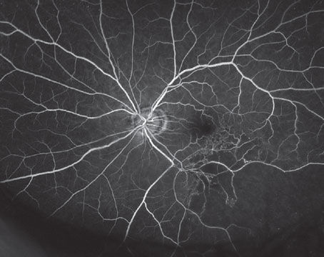
A retrospective, observational study involving 55 patients diagnosed with unilateral Type 3 neovascularization, who later developed neovascularization in the fellow eye, was conducted at the Konyang University College of Medicine in Seoul, South Korea. The study sought to uncover any association between two factors: how much time passed between the previous visit and the visit at which the problem in the fellow eye was discovered (the “examination interval”); and the best-corrected visual acuity and degree of deterioration in that fellow eye when the neovascularization was discovered.
The data showed that there were significant associations between the length of the examination interval and both the BCVA and degree of deterioration in the fellow eye (p=0.005 and p=0.001, respectively). Other findings included:
• The mean interval between the previous visit and the discovery of neovascularization in the fellow eye was 4.8 ±2.2 months (range: 2 to 10 months).
• The time between the initial diagnosis of the first eye and the diagnosis of the fellow eye was 22.7 ±17.5 months.
• Patients were instructed to return to the hospital if they noticed visual deterioration, but only half of the patients recognized deterioration if it was less than three lines, suggesting the importance of monitoring by a health care professional.
The authors state that this result suggests the need for frequent fellow-eye examinations in patients with unilateral Type 3 neovascularization. (The authors note that a high incidence of bilateral involvement is characteristic of Type 3 neovascularization, and preserving the visual acuity in the fellow eye is closely associated with patient quality of life.)
The researchers postulate that a home monitoring system that’s able to detect changes in visual function associated with choroidal neovascularization could provide a practical means to reduce this interval without burdening the patient with extra visits.
Retina 2020;40:7:1255-61.
Kim JH, Kim JW, Kim CG, et al.
MERLOT Turns Sour
Researchers tasked with assessing the efficacy and safety of epimacular brachytherapy for chronic, active, neovascular nAMD report that EMB is ineffective as an adjunctive treatment when combined with anti-VEGF therapy.
The device trial, called the Macular Epiretinal Brachytherapy vs Ranibizumab (Lucentis) Only Treatment (MERLOT), was conducted at 24 National Health Service hospitals across the United Kingdom. Individuals who had nAMD and received intravitreal ranibizumab were enrolled between 2009 and 2012, and were randomized 2:1 and stratified by lens status and angiographic lesion type to receive either EMB plus as-needed ranibizumab, or just as-needed ranibizumab monotherapy. Participants were followed up monthly for 24 months and then assessed at final visits at month 36. Analyses followed the intent-to-treat approach.
Interventions included pars plana vitrectomy with 24 Gy EMB plus as-needed ranibizumab vs. as-needed ranibizumab monotherapy.
Coprimary outcomes were the number of as-needed ranibizumab injections and the mean change in Early Treatment Diabetic Retinopathy Study best-corrected visual acuity, with a noninferiority margin of -5 ETDRS letters.
Secondary outcomes were the percentage of participants losing fewer than 15 ETDRS letters and gaining zero or more, or 15 or more ETDRS letters and the mean change in angiographic total lesion size, choroidal neovascularization size and foveal thickness on optical coherence tomography.
Of 363 participants, 329 (90.6 percent) completed 24 months of follow-up (222 participants in the EMB group and 107 in the ranibizumab group). Here are some of the findings:
• The mean BCVA change was -11.2 ETDRS letters in the EMB group and -1.4 letters in the ranibizumab group, with a difference of 9.8 ETDRS letters.
• In the EMB group, 65.6 percent of participants (160 of 244) lost fewer than 15 letters vs. 86.6 percent (103 of 119) in the ranibizumab group, with a statistically significant difference of 21 percent.
Investigators say that, despite the acceptable safety of EMB, it didn’t reduce the number of ranibizumab injections and was actually associated with worse visual acuity than anti-VEGF treatment alone. They conclude that these results don’t support EMB use as an adjunct treatment for chronic, active nAMD. REVIEW
JAMA Ophthalmol 2020; July 09. [Epub ahead of print].
Jackson TL, Soare C, Petrarca C, et al.