Optos announced the results of a clinical validation study comparing Optos ultra-widefield imaging to Early Treatment Diabetic Retinopathy Study protocol fundus photography, the gold standard for assessing severity of diabetic retinopathy. The study, completed by the Joslin Diabetes Center, was published in the
American Journal of Ophthalmology.
ETDRS protocol seven standard field 30-degree color fundus photography has long been the imaging benchmark for assessing diabetic retinopathy severity. This study reports that the Optos’ ultra-widefield non-dilated optomap images compared favorably with dilated ETDRS photos and dilated retinal examination in determining clinical severity of diabetic retinopathy and diabetic macular edema.
Led by Lloyd Paul Aiello, MD, PhD, researchers at Joslin’s Beetham Eye Institute compared non-dilated ultra-widefield images and ETDRS photos in 103 patients with various severity levels of diabetic retinopathy. The two imaging modalities exactly matched for clinical level of diabetic retinopathy in 84 percent of patients and were within one level of agreement in 91 percent. Sensitivity and specificity of ultra-widefield images for detecting the presence or absence of diabetic retinopathy diagnosed on ETDRS photos were 99 percent and 100 percent.
The study also measured the length of time to capture images using both the Optos device and a traditional digital fundus camera and found that optomap imaging took less than half the time of dilated ETDRS photos not including the time needed to dilate the eyes. Thus, optomap can now allow more efficient imaging while still maintaining the current standards of diabetic retinopathy identification.
Dr Aiello said, “In this study, nonmydriatic ultra-widefield imaging compared favorably with dilated Early Treatment Diabetes Retinopathy Study protocol photography and dilated retinal examination in determining clinical severity of diabetic retinopathy and diabetic macular edema. The additional benefits of easier operation, no pupil dilation and more rapid image acquisition will be significant improvements if these results are confirmed across diverse sites and broader diabetic populations.”
Roy Davis, CEO of Optos, added: “We are extremely pleased with these results which clearly demonstrate that the ultra-widefield optomap technology compares favorably to current imaging standards in assessing diabetic retinopathy. We believe that this study, combined with the increasing body of clinical evidence, demonstrates the benefits of ultra-widefield imaging to clinicians.”
For information, visit
optos.com.
Lock Down Frame Security
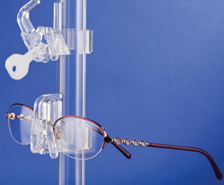 |
Fashion Optical Displays’ new Generation 2 (G2) Locking Frame Support is designed for eye-care professionals who want to deter theft of their inventory. Simply snap each locking frame support onto the system’s crystal clear display tubes. Then place a frame on the G2 support and close the lock to keep frames in place and secure.
With this new snap-and-go lock support, it is easy to unlock and remove the eyewear for customers to try on or to easily rearrange the display. A unique key will unlock all of your G2 locking frame supports. The new G2 supports are easily retrofitted into existing displays and casework.
G2 supports are available as part of the Display Plus Merchandising System that makes it easy to market any frame with its signage display system that directs customers to new designer frames, or to men’s, womens’ and children’s eyewear. Display Plus also offers straight and curved display shelves, mirrors, graphic holders, literature holders, chain and cord holders, etc.
For information visit
fashionoptical.com or call 1 (800) 824-4106.
Iridex Debuts Patient Website
Iridex Corp. has announced the launch of an educational website aimed at patients with diabetic macular edema:
treatmydme.com. The website provides information on MicroPulse Laser Therapy, a new treatment option for DME patients. It explains DME, describes MPLT and addresses patients’ expectations before, during and after treatment.
“The website’s launch will facilitate increased patient awareness and education on MPLT as a viable option for the treatment of DME,” said Sam Mansour, MD, medical director of the Virginia Retina Center in Warrenton, Va., and a clinical professor of ophthalmology at the George Washington University in Washington, D.C. “For patients struggling with DME, it’s important to have easy access to information on the disease and treatment options.”
MPLT is a retina-sparing solution for the treatment of DME. MPLT also can be used in conjunction with drug therapy, allowing complete and optimized management of DME without laser-induced retinal damage, according to Iridex. The laser delivery therapy works by electronically “chopping” the laser emission into trains of microsecond pulses. This enhances the physician’s ability to more precisely control the laser effects on target tissues, offering the potential for ocular treatment with less collateral effects than conventional laser treatments. For information, visit
iridex.com. REVIEW
| Product Research News |
Group Recommendations on Preservatives in Glaucoma Meds
Valeant Ophthalmics announced that the Working Group on Preservatives Toxicity in Glaucoma Medications, an advisory panel of glaucoma treatment experts, recently issued recommendations regarding how to avoid the potentially toxic effects of the preservatives used in many glaucoma medications. The group is sponsored by Valeant Ophthalmics.
The working group discussed the tendency they observed for physicians to deal with ocular surface disease symptoms by adding a steroid or other medication rather than removing the offending agents. Group members disagreed with this “additive” approach and offered “subtractive” alternatives. “The first thing we do is take them off the multiple preserved medications,” said Stephen C. Pflugfelder, MD, Baylor College of Medicine. “My clinic is full of patients that have toxicity, redness or severe lid margin problems. It’s a vicious cycle because the preservative destabilizes the tear film and the whole process just escalates. I found that unless I take them off the offending agent I’m never going to get to the bottom of the problem.”
Reviewing first-line glaucoma therapy options, group members pointed out that prostaglandins were the first choice of most ophthalmologists because of their dosing, efficacy and safety. They recommended a beta blocker such as timolol for additive therapy when required except in cardiovascular and pulmonary patients where contraindicated. Timolol is available in a preservative-free formulation as Timoptic in Ocudose (timolol maleate 0.5%). Timoptic in Ocudose is a topical beta blocker, but is absorbed systemically; therefore the same adverse reactions and contraindications are found with topical administration. “If someone is not at their treatment goal, adding timolol once a day can often times get them to the goal,” reported Don Budenz, MD, MPH, chair of ophthalmology at the University of North Carolina at Chapel Hill. “You can use timolol once a day and get a good effect with similarly low side effects.”
Christophe Baudouin, MD, PhD, University of Paris, reported similar success with the use of beta blockers, reiterated their long clinical history and reported that in Europe, preservative-free beta blocker formulations available since 1997 are experiencing considerable growth. Robert J. Noecker, MD, Ophthalmic Consultants of Connecticut, remarked that in the United States, in contrast, awareness of the benefits of beta blockers and the availability of preservative-free formulations was low among ophthalmologists.
Cornea Publishes Report on LipiFlow System for MGD
The results of a randomized, controlled clinical study of a new treatment for meibomian gland dysfunction and evaporative dry-eye disease, using the LipiFlow Thermal Pulsation System, was reported in
Cornea to provide sustained improvement, on average, in both signs and symptoms. Further, this study demonstrated the clinical utility, safety and effectiveness of LipiFlow in adult patients with MGD and dry-eye symptoms.
The clinical study, supported by TearScience, involved nine U.S.-based investigational centers and 139 patients (278 eyes). It compared results for patients treated with a single LipiFlow treatment to a warm compress control for the treatment of MGD. The average total meibomian gland score for patients who received a single LipiFlow treatment more than doubled from 6.3 ±3.5 at the baseline to 16.7 ±8.7 at four weeks. The increase in the average total meibomian score reflected an improvement in both the quality and quantity of meibomian glands secreting lipids, which keep the water portion of tears from evaporating too quickly, as compared to before the LipiFlow treatment. Two recognized symptom questionnaires were used in the study: the Standard Patient Evaluation of Eye Dryness (SPEED) and the Ocular Surface Disease Index. The average SPEED score for patients who received a single LipiFlow treatment decreased from 14.3 ±4.8 at the baseline to 7.6 ±5.8 at four weeks, demonstrating a mean reduction in dry-eye symptoms. Similarly, the average OSDI score decreased from 32.0 ±20 at baseline to 16.6 ±18.1 at four weeks.
No serious device-related adverse events were reported for the LipiFlow System. Non-serious device-related adverse events included moderate eyelid pain during treatment (three eyes) and transient, moderate eye redness after treatment (one eye). LipiFlow, which applies heat and gentle pressure to a patient’s eyelid, is designed to liquefy and evacuate obstructions in meibomian glands during a 12-minute, in-office procedure. The goal of unblocking the glands is to allow them to resume their natural production of lipids required for a healthy tear film. TearScience manufactures and sells a complete system comprising the LipiFlow and the LipiView Ocular Surface Interferometer, which allows physicians to assess a patient’s tear film.


