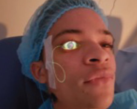
Current surgical options for ocular surface disease range from straightforward traditional procedures such as superficial keratectomy and conjunctival autograft transplantation to more advanced approaches such as limbal and amniotic membrane grafting as well as keratoprosthesis implantation.
OSDs represent a broad range of conditions—from minor corneal scars and localized conjunctival degenerations to more significant limbal stem cell deficiencies and bilateral injury or immune conditions devastating the entire ocular surface. Each case presents an individualized choreographic challenge combining multiple medical and surgical strategies.1 Here the several surgical options that have evolved for OSD management are overviewed.
The 'Big Four' of OSD
Determination of an exact etiologic diagnosis is critical in defining the optimal management strategy. Careful and consistent assessment of the "Big Four" of OSD—blink, tears, sensation and stem cells—is essential to determine appropriate therapy. For example, monitoring the completeness and frequency of the blink plus position and elasticity of the eyelids will indicate if lateral tarsorrhaphy is indicated to avoid corneal exposure or stabilize a "floppy" eyelid.
Carefully observing the ocular surface with flou-rescein or rose bengal vital stains, and performing a Schirmer basic secretion test (with topical anesthesia) may indicate the need for punctal occlusion. Testing corneal sensation with a cotton applicator wisp can suggest if a neurotrophic process requires a therapeutic soft contact lens or even amniotic membrane overlay graft to prevent or manage a persistent epithelial defect.
Remember the dictum of OSD evaluation and management: "Little things mean a lot."
From Simple to Sophisticated
• Superficial keratectomy. For a host of superficial corneal dystrophies and degenerations, ranging from the pannus of epithelial basement membrane dystrophy (EBMD or map-dot-fingerprint) or Salzmann's degeneration to the apical "callus" of keratoconus to even calcific band keratopathy, simple and brief superficial keratectomy, or "super-K", remains remarkably efficient and effective to restore vision and reduce epithelial erosive tendencies.2
If tear function is adequate and corneal sensation is intact, then postoperative surface healing is assured. Although excimer laser phototherapeutic keratectomy has some application, the "low-tech" super-K approach is far more economical, often more effective and hence preferable in most situations. If recurrent erosion should accompany the disorder, as in EBMD, then anterior stromal puncture can readily accompany super-K.
• Conjunctival autograft transplantation. This remains the procedure of choice for a localized inflammatory or degenerative compromise of the conjunctiva and limbus, typically in pterygia but also following cicatricial strabismus, symblepharon or limbal surgical excision.3 The surgical technique remains fundamentally unchanged from the procedure devised at the Massachusetts Eye & Ear Infirmary in the early 1980s with two exceptions: the relatively recent use of tissue adhesives such as Tisseel (Baxter BioSurgery) in place of sutures; and the occasional adjunctive intraoperative application of mitomycin-C.
The rate of success for primary and even recurrent pterygia remains around 95 percent.3
While some surgeons utilize amniotic membrane for this purpose, the time-honored conjunctival autograft remains a most successful and economical approach.
• Limbal autograft transplantation. Also during the 1980s, recognition of stem cells at the limbus as the vital progenitors of corneal epithelium prompted the logical extension of conjunctival autografting to involve the limbal epithelium.4,5,11 For victims of ocular chemical or thermal burns that caused unilateral limbal stem cell deficiencies (LSCD), limbal autograft transplantation proved remarkably curative. Autografted limbal cells are the "gifts that keep on giving." They rehabilitate and permanently restore the ocular surface with no long-term signs of limbal depletion, thereby preventing the "conjunctivalization" of the corneal surface by conjunctiva-derived epithelial cells.6
Thus, the classic limbal autograft remains our preferred approach for uniocular LSCD. This technically straightforward surgery usually suffices for both definitive visual as well as anatomical restoration, because most patients achieve excellent visual recovery without the need for keratoplasty. When stromal scarring significantly impairs vision, limbal autografting also serves as vital preparation for either lamellar or penetrating corneal transplantation. This has been shown to improve keratoplasty success to the 90 percent range, comparable with other favorable prognostic groups.
Variations on the Theme
• Limbal allograft transplantation. For patients with bilateral LSCD due to injury, Stevens-Johnson syndrome or pemphigoid, clearly the near or total lack of limbal tissue suitable for autografting presents greater challenges. Initially investigators devised limbal allograft transplantation, utilizing either living related limbal donors or cadaver eye bank tissue.4 Despite these patients' need for long-term systemic immunosuppression with prednisone, cyclosporine and/or mycophenolate mofetil, limbal allograft transplantation has been somewhat successful, albeit with long-term attrition rates to perhaps 50 percent.
• Ex vivo expansion autografts. Within the past decade, corneal centers with access to cell culture facilities have devised sophisticated techniques for expanding small biopsies (<1 mm) of surviving autologous limbal or oral mucosal tissue grown on amniotic membrane or other suitable substrates. Such ex vivo expansion autografts can then be reintroduced to restore the damaged limbal tissue without the need for immunosuppression.2,7 This clever approach allows the ocular surface to be rehabilitated still in an autograft fashion.
Beyond the Limbus
• Amniotic membrane grafting. In the past 15 years we've also witnessed the reintroduction of human-preserved amniotic membrane (either fresh-frozen or freeze-dried) as a source of collagenous extracellular matrix plus anti-inflammatory, anti-neovascular and epithelialization-stimulating factors. Amniotic membrane grafting has become widely used either as an appropriately sized inlay to fill and heal a stromal defect or as a soft contact lens-sized overlay to heal persistent epithelial defects following herpes simplex or zoster keratitis.3,8 The membrane is either secured with 10-0 vicryl sutures or fibrin adhesive or inserted as a soft contact lens-sized disc adherent to a PMMA ring (ProKera, Biotissue).
The healing properties of amniotic as overlay grafts also improve the outcoomes of limbal grafting, especially for allografts, and high risk keratoplasty, such as with neurotrophic keratopathy. In my own series of more than 30 eyes requiring keratoplasty for various neurotrophic etiologies, many with PEDs, stromal ulceration and neovascularization, the combined use of deep anterior lamellar keratoplasty plus amniotic membrane overlay grafting has substantially improved success to 90 percent.
Continued advances involving this remarkable tissue are ongoing as, for example, a topically applied, lyophilized extract of amniotic membrane has shown excellent promise in recent European trials to successfully heal otherwise unresponsive PEDs.
Prosthetic Solutions
• Keratoprostheses. If patience is a virtue, then persistence is suitable for sainthood, at least as personified by Prof. Claes Dohlman, MD, whose half century quest for a biocompatible keratoprosthesis at MEEI has come to fruition as the Boston K-Pro (to be detailed in a subsequent column). This device has become increasingly successful in eyes with extreme keratitis sicca, exposure or anesthesia, where multiple attempts at ocular surface transplantation and keratoplasty have failed.
A similarly creative biomaterial design also evolving through years of effort is the Boston Ocular Surface Prosthesis, the contribution of Perry Rosenthal, MD, and his associates at the Boston Foundation for Sight. It provides not only a lubricant reservoir spanning the entire ocular surface for eyes with severe keratitis sicca or Stevens-Johnson syndrome and also improves the extreme irregular astigmatism of ectatic corneas affected by keratoconus and pellucid degeneration.
Creative Choreography Is Key
The concept of ocular surface disease as a rubric was devised just 30 years ago.10 Like all paradigm shifts, this has come in its own gestational and evolutionary time. Chemical burns and neurotrophic keratopathy once would have been managed with penetrating keratoplasties, typically doomed to ocular surface failure. Now strategies have been devised specifically to address limbal stem cell restoration and promote and protect the corneal epithelium so that subsequent tissue-specific lamellar keratoplasty enjoys much improved and prolonged visual rehabilitative success.
This approach to OSD management relies on our increasing ability to diagnose disease entities, to recognize their pathogenic mechanisms, and then to select and stage the multiple medical and surgical methodologies to optimize anatomical and functional outcomes. The good fortunate to witness this evolutionary process has for me been a source of great satisfaction and accomplishment as well as encouragement and hope. And most importantly, the saga continues.
Dr. Kenyon is a faculty member at
1. Tseng SC, Tsubota K. Important concepts for treating ocular surface and tear disorders. Am J Ophthalmol 1997;124:825-835.
2. Gangadhar D, Kenyon KR, Wagoner MD. Superficial keratectomy. In: Tasman W, Jaeger EA, eds. Duane's Clinical Ophthalmology.
3. Kenyon KR, Wagoner MD, Hettinger ME. Conjunctival autograft transplantation for advanced and recurrent pterygium. Ophthalmology 1985;92:1461-1470.
4. Kenyon KR, Toda I, Wagoner MD. Ocular surface transplantation. In: Albert DM, Miller JW, Azar DT, Blodi BA, eds. Albert & Jakobiec's Principles & Practice of Ophthalmology.
5. Kenyon KR, Tseng SC. Limbal autograft transplantation for ocular surface disorders. Ophthalmology 1989;96:709-723.
6. Dua HS, Miri A, Said DG. Contemporary limbal stem cell transplantation—a review. Clin Experiment Ophthalmol 2010;38:104-117.
7. Tsubota K, Satake Y, Kaido M, et al. Treatment of severe ocular surface disorders with corneal epithelial stem cell transplantation. New Engl J Med 1999;340:1697-1703.
8. Letko E, Stechschulte SU, Kenyon KR, et al. Amniotic membrane inlay and overlay grafting for corneal epithelial defects and stromal ulcers. Arch Ophthalmol 2001;119:659-663.
9. Lee S, Tseng SCG. Amniotic membrane transplantation for persistent epithelial defects with ulceration. Am J Ophthalmol 1997;123:303-312.
10. Thoft RA, Friend J. The Ocular Surface. Internat Ophthalmol Clinics. Vol 19.
11. Lee H, Kenyon KR. Limbal stem cell transplantation. Contemporary Ophthalmol 2008;7:1-8.


