Presentation
A 56-year-old woman presented with progressive decreased vision in the right eye over the preceding few months. She denied additional ocular complaints such as pain or discomfort. Of note, complete ophthalmic examination in the past year demonstrated unremarkable findings, with the exception of a stable conjunctival nevus.
Medical History
Medical history revealed arthritis, asthma, a benign thyroid nodule, a benign adrenal gland nodule of undetermined histopathology, basal cell carcinoma, chondrosarcoma, a pituitary microadenoma and an auricular glomus jugulare tumor status post proton irradiation. Past ophthalmic history included anatomically narrow angles, which were monitored conservatively, and refractive error. The patient was a non-smoker. Family history revealed age-related macular degeneration, leukemia and multiple sclerosis. Current medications included Allegra, and calcium and vitamin D supplementation.
Examination
On examination, visual acuity with correction was 20/25 OU. Both pupils were equal, round and reactive to light without relative afferent pupillary defect. Intraocular pressure was 15 mmHg OD and 14 mmHg OS. Confrontation visual fields and extraocular motility were full OU. Anterior segment examination was unremarkable aside from a stable conjunctival nevus OS.
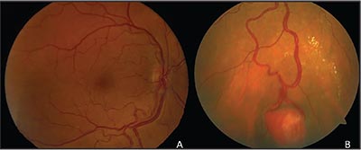 |
| Figure 1. A: Fundus photo of the right eye reveals tortuous, dilated retinal vessels with asymmetric inferior “boxcarring” of the retinal veins. B: A vascular lesion with feeder vessels and surrounding hard exudates was found in the peripheral retina inferiorly. |
On posterior examination, the right eye was noted to have a normal optic nerve appearance and minimal parafoveal drusen. However, the inferior retinal vessels were dilated and slightly tortuous with asymmetric inferior “boxcarring” of the retinal veins (Figure 1A). The far periphery revealed the vessels led to a retinal vascular mass surrounded by exudation (Figure 1B). The posterior evaluation of the left eye revealed mild macular drusen. These features were consistent with the diagnosis of unilateral retinal hemangioblastoma, requiring investigation into whether she had von Hippel Lindau (VHL) disease. Genetic testing for VHL was negative for the mutation.
What is your diagnosis? What further workup would you pursue? Click here for the diagnosis.


