Contact lens wearers who previously experienced contact lens-associated acute red eye had a greater conjunctival response to CL wear when compared with wearers who had no previous inflammatory disease, say researchers from Australia and India. They add that clinical observations were not helpful in predicting wearers who were predisposed to CLARE.
The study enrolled 44 subjects: nine patients who had previously experienced CLARE and nine controls, and 13 subjects who had previously experienced contact lens-induced peripheral ulceration and 13 controls. Controls were matched for age, gender, CL wear experience, and refractive error. All wore bilateral disposable contact lenses (-3.00 D) on one occasion for this study. Contact lens performance and corneal, bulbar and tarsal conjunctival variables were examined upon CL insertion in the evening prior to eye closure, on eye opening in the morning and one, two and four hours later.
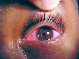 |
| Researchers suggest that a biologic predictor or predisposition to contact-lens associated acute red eye may exist. |
(P < 0.05), bulbar redness (P < 0.05) and conjunctival staining (P < 0.005) compared with controls. Lens fitting and corneal variables were not significantly different at any time point for CLARE subjects. CLPU subjects were not different from matched controls for any variable at any time point. Retrospective analysis of pre-wear variables showed no significant differences between CLARE and control subjects.
Since no clinical, physiological or CL performance factors were revealed in this study as predictors of a subject's susceptibility to develop CLARE or CLPU, researchers suggest that a biologic predictor or predisposition may exist.
(Cornea 2003;22(5):443-447)
Stapleton F, Ramachandran L, Sweeney D, Rao G, Holden B
Gas Proves an Effective Tamponade Over Silicone Oil
C3F8 gas proved to be a more effect-ive tamponade than silicone oil with respect to achieving initial closure of macular holes, says a research group at Duke University Medical Center. In this retrospective, comparative interventional study, 54 eyes of 51 patients underwent pars plana vitrectomy for macular holes. Researchers treated 31 eyes with silicone oil tamponade and 23 eyes with C3F8 tamponade. The patients were demographically similar.
The rate of hole closure after one op-eration with oil tamponade was significantly lower than that with gas tamponade (65 percent vs. 91 percent;
P = 0.022). The percentage of patients undergoing a second operation was significantly higher in the oil group (35 percent vs. 4 percent; P = 0.006).
With reoperations, however, re-searchers found the final rate of hole closure to be similar between the two groups (90 percent for oil vs. 96 percent for gas; P = 0.628). Final visual acuity was better for gas-operated eyes (20/50) than for silicone-operated eyes (20/70; P = 0.047).
(Ophthalmology 2003;110:1170-1174)
Lai J, Stinnett S, McCuen B
Blepharoplasty Reveals Lymphoma
A pair of Houston ophthalmologists recommend histopathologic evaluation of blepharoplasty specimens in cases where the preaponeurotic fat has an unusual appearance, color or consistency at the time of surgery.
In a retrospective review of the medical records of three patients, the researchers discuss the unusual findings at the time of blepharoplasty that led to the diagnosis of orbital lymphoma. All three patients underwent bilateral upper blepharoplasty. His-topathology showed low-grade B-cell lymphocytic lymphoma in all three cases. A postoperative CT scan showed a mass in the lacrimal fossa in one patient. None of the patients had evidence of systemic involvement. Two patients were treated with external beam radiotherapy with good results. One patient recently diagnosed with orbital lymphoma was referred to the oncology clinic for further treatment.
(Ophthal Plast Reconstr Surg 2003;19:316-319)
Arat Y, Boniuk M
Combined Surgery for Proliferative Diabetic Retinopathy
A team of investigators from California and Washington reported that combined phacoemulsification, insertion of a posterior chamber intraocular lens, posterior capsulectomy and pars plana vitrectomy can be used to treat patients with complications resulting from proliferative diabetic retinopathy. They added that combined surgery may prevent a second operation for postvitrectomy cat-aract, allowing for earlier visual rehabilitation.
One of two surgeons performed combined surgery on 223 patients: 153 with vitreous hemorrhage, 58 with traction retinal detachment and 12 with macular traction. Vision increased an average of 4.3 lines. Five percent of patients ex-perienced retinal detachment. Dia-betic macular edema was found in 12 percent of patients after combined surgery. Cystoid macular edema was found in 3 percent. Vitreous hemorrhage requiring another procedure oc-curred in 11 percent. Ten percent required a repeat vitrectomy, 12 for vitreous hemorrhage and 10 for retinal detachment.
Investigators acknowledge the in-creased operating time and surgeon demand made necessary by a combined procedure. They suggest that retinal surgeons may share the surgery with consultation cataract specialists. They don't recommend combined surgery in severely ischemic eyes with rubeosis iridis or in severe traction ret-inal detachments.
(Ophthalmology 2003;110:1335-1339)
Lahey J, Francis R, Kearney J
SLT Effective and Safe for Open-angle Glaucoma
Selective laser trabeculoplasty is effective and safe as a prim-ary treatment for patients with ocular hypertension and open-angle glaucoma, determined researchers from the Goldschleger Eye Institute in Israel. In a prospective, nonrandomized pilot study, 45 eyes of 31 patients underwent SLT as primary treatment. Subjects had open-angle glaucoma or ocular hypertension with intraocular pressure Ž 23 mmHg on two consecutive measurements. Researchers performed visual acuity, slit lamp ex-mination, ophthalmoscopy, gonio-scopy and visual field analysis. They measured IOP at one hour, one day, one week, and one, three, six, 12, 15 and 18 months after surgery. Patients received topical antiglaucoma medications during the follow-up period, if necessary.
Researchers defined success as an IOP reduction of at least 20 percent after SLT. In this study, the mean ±SD decrease in IOP was 7.7 ±
3.5 mmHg (30 percent). The IOP reduction was sustained after SLT, when successful. Forty-three of 45 eyes (93 percent) treated had IOP reductions on two consecutive visits (±2 mmHg).
| Mean Postop IOP and Visual Acuity | |
| Characteristic | Value |
| Follow-up time, mo. | 11±5.3 |
| Final IOP, mmHg | 17.9±2.8 |
| Mean IOP decrease, mmHg | 7.7±3.5 |
| Final VA | 20/30 |
Patients in this study had newly diagnosed untreated glaucoma or eyes with a history of a single topical medication only. Researchers ac-knowledge that this may explain their high rate of success (30 percent IOP reduction) and low incidence and low level of IOP spikes (17.8 percent) one hour after treatment (See Table).
(Arch Ophthalmol 2003;121:957-960)
Melamed S, Ben Simon G, Levkovitch-Verbin H


