It would be nice to be able to aim a device at a LASIK candidate's cornea and have it return an instant verdict of "operate" or "don't operate." Though ophthalmology isn't there yet,
Analyzing Corneal Thickness
Dr. Ambrósio doesn't advocate abandoning topography and pachymetry when screening LASIK candidates. Rather, he feels the addition of measurements from the Oculus Pentacam helps catch patients who may appear normal on traditional indices, and can also avoid branding normal patients abnormal.
Using the Pentacam, Dr. Ambrósio says, "You can evaluate the thinnest point and also its location in Cartesian terms. If you look at the distance between the thinnest point and the center of the cornea—where ultrasound typically measures—and the difference in thickness between the two points, there's a statistical correlation. If the distance is higher, the difference is likely greater. If the point is far away from the center, then the difference in thickness between the points is probably going to be bigger. In 12% of normal corneas, the difference from the central thickness and the thinnest value is over 10 µm."
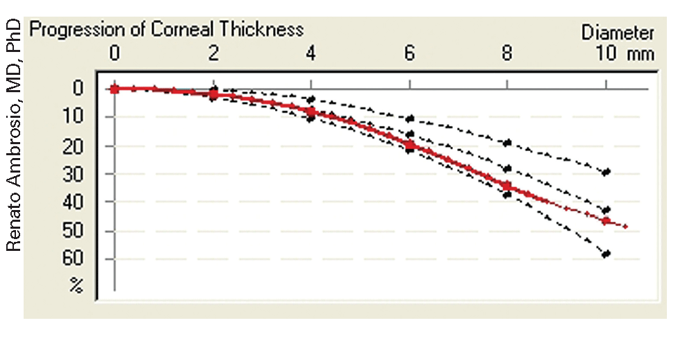
Figure 1. The rapid increase in thickness from the 4.5-mm to 10-mm zones on this Pentacam graph, appearing below as an S-shaped curve in the red line, is a possible sign of abnormality.
The device will also provide a corneal thickness spatial profile (CTSP), which is the average of the thickness values along 22 imaginary circles centered on the thinnest point, with the diameter of the circles increasing in 0.4-mm steps. It also provides the percentage of increase of thickness (PIT), which is the percentage of increase of the average thicknesses in each circle. The CTSP and PIT graphs display the data for the examined eye compared to the 95-percent confidence interval of a normal population.
"The keratoconic cornea has a bigger difference in thickness between the center and the periphery—it gets thicker faster toward the periphery. The normal cornea has a gradual increase that obeys a limit that we can use to define a normal population," says Dr. Ambrósio.
"In a study we performed on 46 eyes with mild to moderate keratoconus and 364 normal eyes, we found statistically significantly different values at all points on the thickness profile and the progression of the increase in thickness," he says.
Dr. Ambrósio says the CTSP and PIT graphs may show early abnormality even before you can identify changes in the curvature maps. "This concept is the basis of a study I am doing with my fellow Marcella Salomão, MD, on cases with very asymmetric keratoconus in which the contralateral eye has a normal corneal surface topography on axial maps," he says. "On the graphs, the line representing the patient's eye may be out of the limits for normality but also exhibit an S shape (See Figure 1) that appears as the diameter increases, meaning that there's a higher progression of the thickness values and some abnormality present."
Corneal Biomechanics
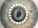
Dr. Ambrósio also adds corneal hysteresis data from the Ocular Response Analyzer into the mix to further screen candidates. The ORA uses a puff of air to get pachymetric and corneal rigidity values. He evaluates the ORA results both numerically and in terms of a morphological graph of the air puff's applanation of the cornea.
Numerically, the device gives two values: corneal hysteresis and the corneal resistance factor. After studying the values in 82 eyes with forme fruste keratoconus and 322 normal eyes, Dr. Ambrósio determined that hysteresis values and CRF values are statistically significantly lower than those in normals (p< 0.05), with an average CH value of 7.81 ±2.25 mmHg and CRF of 7.2 ±2.23 mmHg. In normals, the average value is 10.1 ±1.8 mmHg for CH and 10 ±1.9 mmHg for CRF.
He also studies the waveform signal that is created by the ORA air puff measurement. On the graph, there are three lines: a green line (the air puff pressure) and blue and red lines that represent the filtered and unfiltered infrared signals that peak on applanation. "During the measurement, the air puff applanates the cornea and you get two signal peaks," he explains. "The corneal hysteresis is the difference between peak one and two. CRF is a mathematical calculation based on CH which gives more weight to the first applanation pressure. If a cornea is strong, it will absorb more energy, and therefore the difference will be higher and it will take longer for the cornea to go back because it's stiffer. If it's weaker, the difference between point one and two will be less. A very important sign of a weak cornea will be a red line that, after the second peak, goes down and up again, with a little peak after that." (See Figure 2, below.)
Case Studies
Dr. Ambrósio gives two examples in which the enhanced ectasia indices shed light on certain cases.
In the first case, a 28-year-old developed progressive ectasia seven years after LASIK in one eye, even though there were no identifiable risk factors preop. Before surgery, the central corneal thickness was 528 µm, the flap thickness was 159 µm and there was a 270-µm residual stromal bed, which is within the usual 250-µm guideline followed by many surgeons. Topography was normal, and the refraction was -6 D OD and -5.75 D OS.
The unoperated right eye has actually remained stable over time and would appear to be a good candidate for LASIK by conventional screening. However, when the expanded indices are applied to the unoperated eye, red flags fly. On the Pentacam, his CTSP and PIT graphs are borderline. On the ORA, his corneal hysteresis and corneal resistance values actually show a high susceptibility for ectasia. If these indices had been applied to the first eye before it underwent LASIK, it's possible that the ectasia might have been avoided.
Dr. Ambrósio says the following case illustrates the opposite possibility: Validating the decision to do LASIK on a patient who might be branded a bad candidate due to slightly unusual topography.
In this case, the patient was a 23-year-old, fairly high myope with mild topographic asymmetry that might be considered suspicious. However, when Dr. Ambrósio performed his enhanced indices, the picture became clearer. "The patient had a normal thickness profile and a very good biomechanical profile—there was a value of over 12 mmHg for both hysteresis and her corneal resistance factor," he recalls, "so it's very much a good cornea. We did LASIK and she's doing very well as of a year postop.
"This patient is an example that shows we may have avoided LASIK based only on topography, which might not have been specific enough to detect some aspects of the eye."