An 84-year-old Caucasian female presented for evaluation of a right iris lesion with associated symptoms of glare and light sensitivity for the preceding three months. She had had a similar occurrence approximately two years earlier, and vaguely recalled that it resolved after treatment with a laser procedure. The patient was referred to the Ocular Oncology Service at Wills Eye Hospital for further evaluation and management.
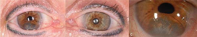 |
| Figure 1. External photography of the right eye (A) shows a cystic iris lesion inferiorly measuring 10 mm x 6 mm, as well as blepharopigmentation in the upper and lower eyelids. The left eye (B) reveals similar blepharopigmentation. A magnified view of the right eye (C) depicts the cystic lesion protruding into the anterior chamber and distorting the pupil. |
Medical History
Her past ocular history was notable for previous uncomplicated cataract surgery in both eyes, as well a giant retinal tear with detachment in the right eye that required surgical repair. She had bilateral blepharopigmentation (eyeliner tattoos). Her medical history was notable only for hypertension, controlled with atenolol. She had no known allergies, and her family and social history were unremarkable.
Examination
On ocular examination her best corrected visual acuity was 20/60 OD and 20/30 OS. Intraocular pressures were
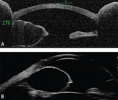 |
| Figure 2. Anterior segment optical coherence tomography of the right eye (A) shows an iris cyst originating from the stroma and occupying the anterior chamber. Ultrasound biomicroscopy of the right eye (B) confirms an iris cystic lesion extending anteriorly with associated endothelial touch. |
Anterior segment optical coherence tomography was performed OD and demonstrated a thin-walled, fluid-filled, cystic lesion in the iris stroma distorting the iris both anteriorly and posteriorly (Figure 2A). Ultrasound biomicroscopy OD confirmed the cystic iris stromal lesion with endothelial touch (Figure 2B).
Click here for the Diagnosis and Discussion.


