Digital camera makers have done a good job of equating a camera's megapixel rating with its quality relative to other cameras, leading consumers to go after more and more megapixels. However, experts in the field of digital fundus photography say these megapixel wars can cause you to buy more camera than you actually need for your clinic, or neglect other aspects of the imaging system that are just as important when it comes to creating a good image. Here's what's important in a fundus camera system and where megapixels fit in the mix.
Get a Sense of Sensors
In addition to the megapixel capacity of a camera's light sensor, the actual size of the sensor and the quality control involved with the sensor's construction play a part in getting a good image.
"There are consumer-grade, 5-megapixel sensors and then there are scientific or medical-grade 5-megapixel sensors that cost four to five times as much," says Lon Dowell, who was once the imaging director for a large retina practice and is now director of marketing for imaging products at Topcon. "How can this be? It's the quality of the image sensor itself, the quality of the lenses that focus the image and such factors as whether or not clean rooms were used to assemble and test the sensor and so forth."
The size of the sensor also plays a part, say experts. "It's really more than megapixels. It has to do with sensor size," says Frank Wood, marketing manager for Nidek. "Our goal is to get the image quality as close to 35-mm film photography as possible. If the image isn't at a one-to-one ratio, then the image is digitally altered and lacks the natural color needed for exact diagnosis. The higher the megapixels, plus the larger the sensor, the more accurate the image."
Mr. Dowell says the larger the sensor, the more sensitive the camera is to light. "The larger the pixels the better, because the sensor then has more light-gathering components," he says. "In ophthalmology, it's especially important to have what I call a high-res sensor, but it's almost more important to have a highly sensitive sensor. For instance, say you're doing a fluorescein angiogram in which you place optical filters in the optical path of the fundus camera. These filters will cut down on the light that reaches the sensor, so you want the sensor to be as sensitive as possible to the light spectrum you're introducing to it."
Ron Kaiser, national sales manager for Canon, says that even though there are benefits to larger sensors, there may be trade-offs, as well. "If you go to a bigger sensor, such as full-frame [35-mm size], you have larger pixels that help gather more light," he says. "You can then use a lower flash. However, you may lose some detail in the image because the micron size of the smallest feature the camera can resolve won't be as fine. For example, the Canon 5D camera has a micron size of 6.8, while the 50D and 40D come in at around 4.3 µm, meaning the resolving power is better for what you're actually getting on the sensor."

Mr. Kaiser says another factor to inquire about when shopping for a fundus camera is how the technology in the sensor is used to get rid of "noise," or variations in the color values of individual pixels, especially in areas supposed to be black. These variances manifest on a digital image as small speckles. "Depending on how the camera processes an image, it will either produce or reduce noise," explains Mr. Kaiser. "Both outcomes can cause issues on the final image you see. So, this function has to be adjusted correctly."
How Many Pixels Do You Need?
Even though cameras' megapixel ratings continue to climb, it turns out you only need a modest amount of sensor resolution to visualize common retinal features, and, due to the inherent mismatch of a rectangular sensor imaging a round fundus, you don't even use all the pixels a camera has.
"Diabetes microaneurysms need at least a 3-megapixel sensor," says Mr. Kaiser. "Some finer things need 6 megapixels. Also, it's important to keep in mind that no ophthalmologist will use the full size of the sensor, and will, instead, lose about a third or more of the megapixels because the round fundus image only populates about 50 to 60 percent of the sensor size. So if you have a 6-megapixel camera back, for example, you may use 2.8 megapixels for the retina image." Topcon's Mr. Dowell estimates an 8-MP sensor is more than enough for 99 percent of cases.
Another factor that may make it possible to have too many pixels is the limit of the human eye to appreciate such high-resolution images.
"The human eye has a limited ability to resolve spatial resolution," says Mr. Dowell. "So, at some point, the resolution will reach the level at which you can't tell the difference between 3-, 4-, 8- and 11-megapixel images. I have proven this time and again at trade shows. At our booth, I'll show someone a 4-megapixel image and an 11-megapixel image of the same eye and the person won't be able to tell the difference … It would matter if you blow the image up to a large size, but, in a medical setting, when using a 24-inch monitor, doctors aren't enlarging images to the degree that they'd see actual pixel detail on the screen. In my estimation, more pixels just mean more bragging rights."
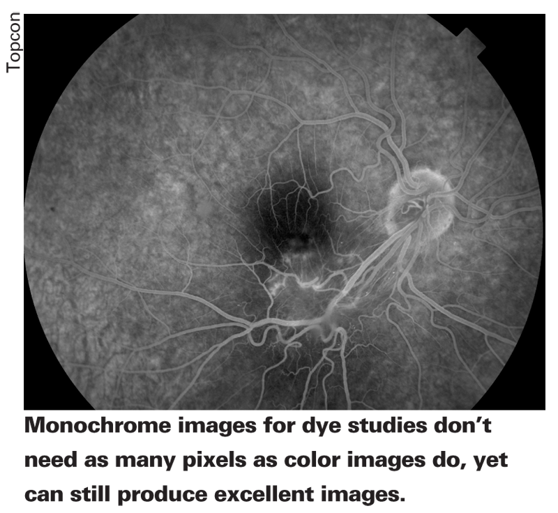
Also, if you rely heavily on digital images of fluorescein angiography, which are presented in monochrome, then you don't need an abundance of megapixels. This is because black-and-white images can use fewer pixels and still be razor sharp. "A monochrome sensor can perform a good enlargement to view fine detail with 1.5 megapixels," says Mr. Dowell.
Monitors and the Human Factor
Another reason a user may not need a great number of megapixels is that his monitor may not be capable of displaying all of them.
"If you have an 8-, 10- or 12-megapixel device, for instance, and you want your monitor to show all of that resolution, the monitor will cost $12,000 to $13,000," says Mr. Kaiser.
"A 6-megapixel monitor is closer to $2,000, but many physicians don't want to purchase such a device for every exam room. At trade shows, we use 21-inch monitors that display around 4 megapixels and cost around $1,000. For everyday use in a practice, most physicians are running monitors with resolutions in the neighborhood of 1200 x 760 or 1800 x 1200." Mr. Dowell recommends sticking with higher-end name brands for your monitor and getting the lowest "dot pitch," or the distance between pixels, that you can.
A lower dot pitch number, measured in millimeters, equals a sharper overall image.
The other factor that comes into play with digital fundus photography is the device's focusing ability, which also often involves the skill of the operator. "The higher-end mydriatic camera in which megapixels will be more important is usually used in a retinal specialist's office or the office of a comprehensive ophthalmologist who's interested in retinal disease," says Mr. Dowell. "This instrument needs a highly skilled technician to look through the fundus camera and focus on fine details of the retina. If they don't perform well, they're going to get a blurry image. So, if you have a patient with a cataract, a lower-skilled tech but a sophisticated system, you'll still get a bad image. However, if you have a skilled tech but a somewhat lower-resolution system, you'll probably get a pretty good image. He or she will know how to shoot around the cataract. Though some of the image will be out of focus, he will be able to capture the pertinent areas of the retina in clear focus.
"I think that, overall, imaging sensors are all very good," adds Mr. Dowell. "There's really no need for 10 megapixels or more. Companies offer them because some camera users want to have bragging rights, but, clinically, it's not signficant."


